Kidney Res Clin Pract > Epub ahead of print
Nan, Piao, Jin, Chung, Yang, and Li: Pathogenesis and management of renal fibrosis induced by unilateral ureteral obstruction

Abstract

Regardless of the underlying etiology, renal fibrosis is the final histological outcome of progressive kidney disease. Unilateral ureteral obstruction (UUO) is an ideal and reproducible experimental rodent model of renal fibrosis, which is characterized by tubulointerstitial inflammatory responses, accumulation of extracellular matrix, tubular dilatation and atrophy, and fibrosis. The magnitude of UUO-induced renal fibrosis is experimentally manipulated by the species chosen, animal age, and the severity and duration of the obstruction, while relief of the obstruction allows the animal to recover from fibrosis. The pathogenesis of renal fibrosis is complex and multifactorial and is orchestrated by activation of renin-angiotensin system (RAS), oxidative stress, inflammatory response, transforming growth factor beta 1-Smad pathway, activated myofibroblasts, cell death (apoptosis, autophagy, ferroptosis, and necroptosis), destruction of intracellular organelles, and signaling pathway. The current therapeutic approaches have limited efficacy. Inhibition of RAS and use of antioxidants and antidiabetic drugs, such as inhibitors of sodium-glucose cotransporter 2 and dipeptidyl peptidase-4, have recently gained attention as therapeutic strategies to prevent renal scarring. This literature review highlights the state of the art regarding the molecular mechanisms relevant to the management of renal fibrosis caused by UUO.

Introduction

Chronic kidney disease (CKD) is a serious public health problem worldwide [1]. Despite strict glycemic control and the use of antiproteinuric drugs (e.g., angiotensin-converting enzyme inhibitors [ACEIs] and angiotensin receptor blockers [ARBs]), the incidence of CKD continues to increase because of the increasing prevalence of diabetes mellitus, hypertension, and obesity, as well as population aging [2]. The prevalence of CKD has been reported as 14% in the United States [3], 10.8% in China [4], and 8.2% in South Korea [5]. A systematic review and meta-analysis of observational studies reported that the global mean prevalence of stage 3 to 5 CKD is 10.6% and that of stage 5 is 13.4% [6]. The recommended management of CKD includes nutritional therapy, treatment of albuminuria, cardiovascular risk reduction, avoidance of potential nephrotoxins, adjustments to the dosage of drugs such as antibiotics, and management of complications, including hyperkalemia, anemia, and metabolic acidosis [1].
As a common clinical cause of renal injury, obstructive nephropathy is usually caused by urinary tract stones, benign prostatic hyperplasia, and pelvic or ureteral tumors. It can occur at all ages from infancy to old age and eventually leads to renal fibrosis. In addition, it may lead to acute kidney injury (AKI) or CKD. Obstruction of the urinary tract decreases renal blood flow and the glomerular filtration rate (GFR), and hinders reabsorption of solutes and water, urine concentrate, and excretion of hydrogen and potassium. Renal fibrosis is the final pathway and histological outcome of most forms of progressive kidney disease, including long-term obstructive nephropathy. Unilateral ureteral obstruction (UUO) is an ideal reproducible experimental rodent model of progressive renal fibrosis [7]. Although the UUO model enables the study of the different stages of renal fibrosis development in an accelerated manner, the precise mechanisms underlying UUO-induced renal fibrosis are not fully understood. Oxidative stress plays a major role in tubular and/or cellular injury and initiates the overproduction of reactive oxygen species (ROS), interstitial inflammatory cell infiltration, activation of fibroblasts, upregulation of transforming growth factor beta 1 (TGF-β1), cell death (apoptosis, autophagy, ferroptosis, and necroptosis), deposition of the extracellular matrix (ECM), and renal fibrosis [8]. Oxidative stress also evokes mitochondrial damage and prolonged endoplasmic reticulum (ER) stress, both of which lead to apoptosis [9].
This review article summarizes the current understanding of the pathogenesis of UUO-associated renal fibrosis and discusses the current literature on the prevention and delay of the progression of this disease.

The unilateral ureteral obstruction model

In general, the UUO model is initiated in rodent animals, mainly rats and mice, by ligation of the left ureter, which leads to ureteral obstruction, hence the term “ureteral obstruction.” The reason for choosing the left ureter may be related to the lower position of the left kidney, which avoids damaging the liver. The ureter in the contralateral (right) kidney is not ligated and is called the nonobstructed or unobstructed kidney. Male animals are preferred because the presence of female reproductive organs complicates the surgical procedure. It is important that the animal’s body temperature is maintained and the kidney is kept moist during the surgery. In this procedure, the animal is anesthetized, and an incision is made in the flank or middle abdomen to expose the kidney and ureter. The left ureter is ligated with silk thread and the incision is sutured. The types of ligation are single ligation, double ligation, and two-place ligation and cut. Sham surgery is performed for the UUO procedure without ligation of the left ureter. The overall treatment protocol for UUO duration ranges from 3 days to 6 months, and most studies collect data on day 7 or 14. Regardless of the kidney under UUO, the contralateral kidney illustrates several alterations during chronic UUO, such as enlarged interstitial spaces and impaired mitochondrial function (after 14 days) [10], dilated distal tubules, cast formation, collagen deposition, and fibrosis (after 6 months) [11,12]. Interestingly, at day 7 and even day 14, the UUO does not influence renal function (e.g., serum creatinine, blood urea nitrogen, and cystatin C) [13,14]. This may be associated with the compensatory function of the contralateral kidney at the early stage of UUO. The various UUO animal models reported in the literature are listed in Table 1 [8,9,11,12,1522].

Manifestations of unilateral ureteral obstruction-induced kidney pathology

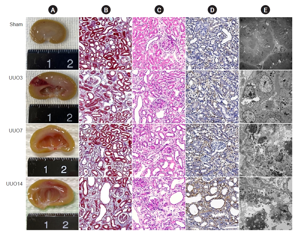
Images of the gross findings show that UUO results in enlargement of the kidney with hydronephrosis and pyelectasis, destruction of the renal pelvis, and an extremely thin kidney cortex (Fig. 1A) [7,23]. Pathology staining and transmission electron microscopy show that the major changes are confined to the tubulointerstitium and are characterized by necrosis, tubular vacuolization and atrophy, swelling of the tubular epithelium and interstitium, inflammatory cell influx, and fibrosis (Fig. 1BD) [14,24].

Pathogenesis of unilateral ureteral obstruction-induced renal fibrosis

Renin-angiotensin system

Blockage of urine flow caused by UUO increases the hydrostatic pressure and impacts feedback to the distal and proximal tubules, which leads to mechanical stretching that damages the tubular cells and decreases the GFR. Both short-term UUO (as early as 6 hours) [25] and prolonged UUO (5 weeks) [26] activate the components of the renin-angiotensin system (RAS), such as expression of renin messenger RNA, ACE activity, and Ang II content, which contribute to renal fibrosis. These effects can be reversed by ARBs [27,28] or ACEIs [29]. Recently, our laboratory found that the angiotensin receptor neprilysin inhibitor LCZ696 alleviates renal fibrosis via ASK1-JNK-p38 MAPK-mediated apoptosis in the UUO model [30].

Oxidative stress

Oxidative stress plays a pivotal role in a wide array of kidney diseases through the overproduction of ROS [31]. In normal conditions, ROS production is strictly governed by the antioxidant system and ROS act as second messengers that help to regulate biological activity through oxidation of redox-sensitive proteins [32]. In response to pathological stimuli, the imbalance between the oxidant and antioxidant enzymes leads to an increase in the release of proteins involved in oxidation, which contributes to renal scarring [33]. Emerging evidence shows that oxidative stress contributes to renal fibrosis even in the early stage of UUO. Oxidative stress is manifested as lower activities of antioxidant enzymes such as catalase, superoxide dismutase (SOD), glutathione, and glutathione peroxidase in obstructed kidneys compared with sham-operated kidneys [34] and increased activity of oxidant enzymes, including nicotinamide adenine dinucleotide phosphate (NADPH) oxidases (NOXs), N(epsilon)-carboxymethyl-lysine, 8-hydroxydeoxyguanosine (8-OHDG), malondialdehyde, and 4-hydroxynonenal [24,35]. Pharmacological administration of antioxidant agents [14] or genetic knockout of cyclooxygenase 2 [36] or thioredoxin-interacting protein [37] alleviates renal fibrosis in UUO models.

Interstitial inflammation

Interstitial inflammation precedes ongoing fibrosis and is the predominant feature of renal fibrosis in UUO. The inflammatory response triggers a self-protective response against deleterious stimuli but sustained inflammation drives deposition of the ECM in the interstitium, which leads to irreversible fibrosis [38]. Numerous proinflammatory and profibrotic cytokines produced by renal tubular cells, myofibroblasts, and infiltrating macrophages are involved in the inflammatory response in UUO. Of these, macrophages are the major source of chemokines and cytokines such as monocyte chemoattractant protein 1, chemokine receptors, adhesion molecules, and TGF-β1 [39]. Chemokines and TGF-β1 also recruit macrophages, and this leads to a vicious cycle of inflammation [40]. Deficiency in NLRP3 or interleukin 36 (IL-36) attenuates renal fibrosis, as evidenced by blockade of ECM deposition, downregulation of NLRP3, pro-IL-18, pro-IL-1, and pro-caspase-1, and decreased inflammatory cell infiltration in UUO [41].

Fibroblast activation

Myofibroblasts belong to a heterogeneous superfamily derived from a variety of origins, including the epithelium through epithelial-to-mesenchymal transition, endothelia through endothelial-to-mesenchymal transition, local fibroblast or pericyte proliferation, and the macrophage-to-myofibroblast transition [42]. During renal fibrosis associated with UUO, activation of myofibroblasts is a key factor in the role of matrix-secreting cell types in ECM accumulation. About half of the myofibroblasts are created via the proliferation of local residential fibroblasts, 35% of nonproliferating myofibroblasts are created by differentiation of cells in the bone marrow, 10% come from the endothelial-to-mesenchymal transition program, and 5% come from the epithelial-to-mesenchymal transition [43]. TGF-β1, a member of the TGF-β superfamily, plays a critical role in this process via Smad-dependent and -independent signaling pathways [44].

Programmed cell death

Apoptosis (type I programmed cell death) is an active mechanism of cell clearance and plays an essential role in cell number regulation during development and organ homeostasis, and following insults [45]. In physiological conditions, apoptosis may be beneficial but can become deleterious if enough resident cells are lost [14]. The apoptotic program is classified according to the extrinsic and intrinsic pathways. Upon extrinsic stimulation, caspase-8 is activated by the ligation of death receptors that further activate downstream caspases by the extrinsic pathway. In the intrinsic (mitochondrial) pathway, proapoptotic Bax and Bak form pores in the mitochondrial outer membrane, which cause the release of apoptogenic factors (cytochrome C) and lead to stimulation of conformational modifications in the adapter protein Apaf-1 and subsequent activation of caspase-9 [46]. Conversely, Bcl-2 and Bcl-xL hinder pore formation via binding to Bax and Bak [47]. Overwhelming evidence has confirmed a pathological role of apoptosis in various kidney diseases as well as UUO [48].
Autophagy (type II programmed cell death) is an evolutionarily conserved, lysosome-mediated cellular process of degradation of damaged organelles, protein aggregates, and other macromolecules in the cytoplasm, and is involved in the regulation of cell death and proliferation [49]. The process of autophagy entails several consecutive procedures that produce autophagosomes and autophagolysosomes, and this process is controlled by a series of genes, such as autophagy-related Atg and LC3 [50]. Abnormalities in autophagy are implicated in a variety of kidney diseases [51]. In UUO, genetic ablation of Atg7 in distal tubules or pharmacological blockade of autophagy aggravates renal fibrosis, which suggests a protective role for autophagy [52]. However, this self-cannibalistic potential may be deleterious if excessive renal cells are lost. Livingston et al. [53] reported that persistent activation of autophagy worsens renal fibrosis in UUO mice with proximal tubule knockout of Atg7. Autophagy is also related to the upregulation of connective tissue growth factor and collagens, myofibroblast differentiation, apoptosis, mitochondrial dysfunction, and fibrosis [54]. Given its diverse actions, autophagy may serve a dual role as either a protector or contributor to kidney disease even within the same condition.

Intracellular organelles

The integrity of mitochondria and ER fitness play a pivotal role in organ pathophysiology [33]. In the kidney, mitochondria are more susceptible to oxidative stress because of arteriovenous oxygen shunting and their high oxygen consumption [55]. In kidney injury, oxidative stress causes mitochondrial ROS overproduction, which generates oxidative stress, alters the cell redox-sensitive signaling pathways, and results in inflammation and apoptotic cell death [31].
Sustained ER stress is cytotoxic to renal cells [33] and mitochondrial damage and ER stress are involved in the development of renal fibrosis in the UUO model [24,33]. In images generated using transmission electron microscopy, it is apparent that UUO destroys the mitochondrial architecture, as manifested by reductions in the number and size of mitochondria, vacuolization, mitochondrial deformation, and mitochondria divided into two daughter organelles (Fig. 2A). UUO is also closely associated with degranulation of ribosomes, disconnected and dilated cisternae, and peroxisome vacuolization in the rough ER, although the smooth ER remains almost normal in structure (Fig. 2B).

Necroinflammation

Necroptosis is a genetically regulated form of cell death controlled by receptor-interacting protein kinases 1 and 3 (RIP1 and RIP3) and downstream substrate pseudokinase mixed lineage kinase domain like (MLKL) [56]. Necrotized cells release danger-associated molecular patterns, which stimulate innate immunity to provoke the sterile inflammatory response via the ruptured plasma membrane. The sterile inflammation in turn aggravates necroptosis via tumor necrosis factor alpha (TNF-α) or interferon [57,58]. Therefore, the term “necroinflammation” refers to the reciprocal boosting of necrosis and sterile inflammation in an autoamplification loop [57,59]. Studies have suggested that RIP1-RIP3-MLKL-mediated necroinflammation is involved in the early stage of AKI [60]. However, Yi and OuYang [61] and Chen et al. [62] reported that necroinflammation participates in renal injury within 6 weeks in diabetic nephropathy (DN) and in the progression from AKI to CKD at 12 weeks. Of particular interest is that RIP3-MLKL signaling accelerates the inflammatory response by activating the NLRP3 inflammasome and thereby regulates the maturation of pro-IL-1β and pro-IL-18 [56]. We and others have reported that blockade of necroptosis with a specific RIP inhibitor (necrostatin-1 or GSK872) or RIP3 deficiency attenuates inflammation and fibrosis in UUO [13,24].

Renal fibrosis

Renal fibrosis is the final pathway of CKD progression that leads to end-stage renal disease requiring renal replacement therapy. Expansion and collagen fiber deposition within the tubulointerstitium are typical hallmarks of renal fibrosis. The major components of the ECM are collagens I, III, and IV, and fibronectin, and TGF-β1 may play an essential role. Consistent with the pleiotropic activities of this growth factor, emerging evidence suggests that TGF-β1 participates in the pathogenesis of a wide range of kidney diseases characterized by glomerulosclerosis and tubulointerstitial fibrosis [63]. In tissues, TGF-β is secreted as a biologically inactive complex requiring in vivo activation. The TGF-β1 inducible gene-h3 (βig-h3) encodes a matrix protein that is regulated by TGF-β. βig-h3 is regarded as an indicator of TGF-β1 bioactivity in renal injury and its expression is thought to indicate the extent of interstitial injury in the human [64] and rat kidney [65].

Congenital and epigenetic mechanisms

In addition to the above-mentioned factors, congenital and epigenetic mechanisms should also be considered. In clinical practice, congenital obstructive uropathy is a leading cause of pediatric renal failure associated with kidney dysplasia in infants and children. Most cases of congenital obstructive uropathy resulting from the posterior urethral valves are tightly linked to varying levels of injury to the bladder, ureter, and kidney. This clinical dilemma manifests as a reduction in nephrogenesis, glomerular maturation, and tubular cellular proliferation accompanied by a greater compensatory growth of the opposite kidney [66]. The mechanisms underlying congenital obstructive uropathy remain incompletely defined but may be associated with combinations of biomechanical signal transduction, growth factor expression, and responses of specific renal autoregulatory mechanisms. Therefore, numerous animal models of congenital obstructive nephropathy have been developed, such as the chick embryo, fetal rabbit, and opossum. Moreover, epigenetic mechanism refers to heritable alterations in target gene expression via post-translational modifications of protein complexes, namely histones, or DNA methylation without altering the underlying genetic DNA sequences. The post-translation protein modifications include acetylation, methylation, phosphorylation, sumoylation, ubiquitinylation, carbonylation and glycosylation, and DNA methylation. Of these, two major types of epigenetic regulations that have been implicated in the pathogenesis of kidney diseases are histone acetylation and DNA methylation. A growing body of evidence suggests that epigenetic modifications in gene expression participate in the process of renal fibrosis, inflammation, and epithelial-to-mesenchymal transition in CKD. Pharmacologic inhibition of histone deacetylase attenuates renal fibrogenesis by amelioration of inflammation and ECM gene transcription and restoration of bone morphogenetic protein 7 (BMP7) and Klotho protein in UUO [67,68]. By detecting their aberrant expression, epigenetic modifiers might serve as potential biomarkers for diagnosing UUO-related kidney disease, and they may also be used as therapeutic targets for the management of these diseases.
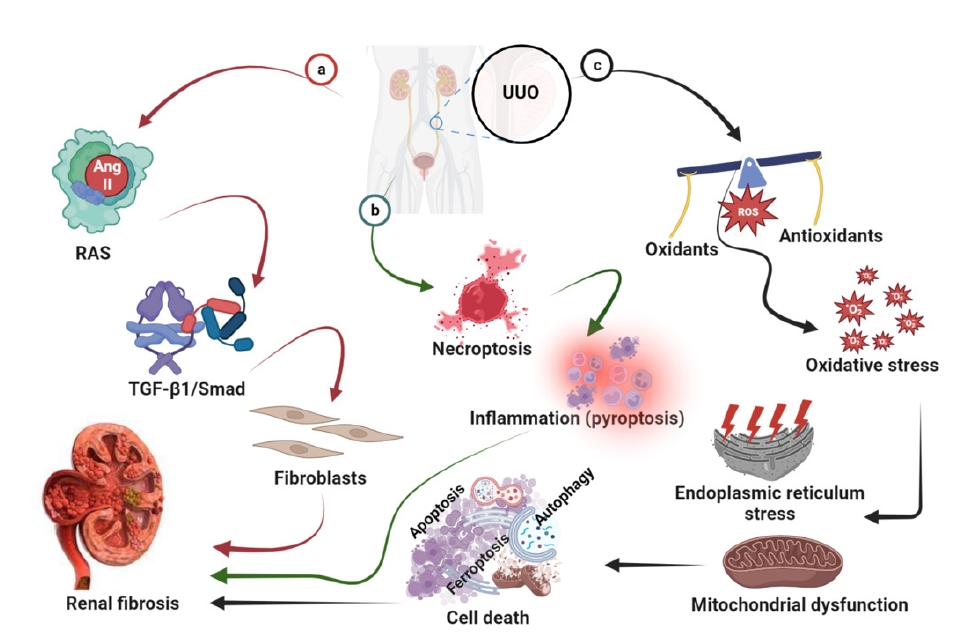
A diagram of the pathogenesis of UUO-induced renal fibrosis is shown in Fig. 3.

Management of unilateral ureteral obstruction-induced renal fibrosis

LCZ696

LCZ696 (valsartan/sacubitril) is a dual-acting angiotensin receptor neprilysin inhibitor. It is one of the new drug classes used for the management of hypertension and heart failure (HF) with reduced ejection fraction. The drug combines the angiotensin receptor blocker, valsartan, and the neprilysin inhibitor prodrug, sacubitril, in a 1:1 ratio in a sodium supermolecular complex [69]. Dual inhibition of neutral endopeptidase and Ang II by LCZ696 confers a better effect than each alone because it avoids reactivation of the RAS and angioedema [70]. The large PARADIGM-HF clinical trial reported that LCZ696 treatment caused a slower rate of decline in the estimated GFR, and improved cardiovascular outcomes compared with enalapril in patients with HF and CKD [71]. This concept is supported by animal studies of DN [72], subtotal nephrectomy [73], and UUO [30].

Vitamin E

Vitamin E is a fat-soluble vitamin also called alpha-tocopherol. Vitamin E has antioxidant potential and maintains intracellular integrity by protecting cells from oxidative stress [74]. In UUO, vitamin E ameliorates the progression of renal fibrosis by suppressing the TGF-β-Smad2/3 signaling pathway [75].

Curcumin

Curcumin is a natural phenolic compound extracted from the rhizome of the plant Curcuma longa (Zingiberaceae) that exhibits various biological activities. Previous studies have reported that curcumin improves renal dysfunction through regulation of the immune system, suppression of inflammatory mediators, oxygen free radicals, and apoptosis, and preservation of mitochondrial integrity [76]. A systematic review and meta-analysis of clinical trials found that curcumin supplementation has potential as a treatment for CKD [77] and diabetic kidney disease [78]. These beneficial effects of curcumin have also been reported in animal models of ischemia-reperfusion injury [79], DN [80], and UUO [81].

N-Acetylcysteine

N-acetylcysteine (NAC) is a modified amino acid that can reverse the toxic effects of acetaminophen overdose and prevent hepatic injury. The amino acid cysteine has a thiol side chain that can facilitate redox reactions and thereby exerts antioxidant activity [82]. Importantly, cysteine is needed for the synthesis of glutathione, which is a key intracellular antioxidant that protects against free radicals and other intracellular toxins [83]. Because of this activity, NAC may exert anti-inflammatory and antifibrotic actions in a variety of kidney diseases [84]. Shen et al. [85] reported that NAC attenuates the expression of collagen III and TNF, and renal fibrosis in UUO.

Sodium-glucose cotransporter 2 inhibitors

Sodium-glucose cotransporter 2 (SGLT2) inhibitors have been approved for the treatment of all stages of type 2 diabetes mellitus (T2DM). This class of drug directly blocks SGLT2 and thereby inhibits renal glucose reabsorption, boosts urinary glucose excretion, and effectively lowers hyperglycemia [86]. SGLT2 inhibitors afford better blood glucose control than other antidiabetic agents and have several advantages such as a low incidence of hypoglycemic episodes and lower body weight, and blood pressure [87]. Clinical trials have confirmed that SGLT2 inhibitors confer renoprotection and cardioprotection in patients with T2DM [88]. We have shown that dapagliflozin ameliorates renal fibrosis by inhibiting RIP1-RIP3-MLKL-mediated necroinflammation via Wnt3α/β-catenin/GSK-3β signaling in UUO rats [13].

Dipeptidyl peptidase 4 inhibitors

The first dipeptidyl peptidase 4 (DPP4) inhibitor was approved for the management of diabetes in 2006. This drug reduces blood glucose concentration primarily by impeding the degradation of glucagon-like peptide-1 and -2 (GLP-1 and GLP-2) and glucose-dependent insulinotropic peptide [89]. DPP4 is expressed predominantly in the glomeruli and the brush border of the S1 to S3 segments of the proximal tubule [90], whose actions include promotion of the absorption of cleaved dipeptides and regulation of the function of the sodium-hydrogen exchanger 3 in the proximal tubules. Inhibition of DPP4 may increase its bioavailability, thereby prolonging the half-life of insulin action. DPP4 inhibitors have been approved for treating T2DM as a monotherapy, add-on therapy, or combination therapy with other antidiabetic agents. Beyond glucose reduction, DPP4 inhibitors elicit renoprotective effects through either a GLP-1-independent proteolytic function or a nonproteolytic DPP4 function [91]. In a mouse model of UUO, the DPP4 inhibitor, LC15-0444, significantly decreased albuminuria, urinary excretion of 8-isoprostane, synthesis of several proinflammatory and profibrotic molecules, and renal fibrosis [92]. These findings suggest that DPP4 inhibitors have pleiotropic effects.

L-Carnitine

The quaternary ammonium compound L-carnitine (LC, L-β-hydroxy-γ-N-trimethylaminobutyric acid) acts as a key cofactor for the β-oxidation of fatty acids by facilitating the transport of long-chain fatty acids in the form of acylcarnitine esters across the mitochondrial membrane [93]. The derivatives of β-oxidation (two carbon molecules) are used by the Krebs cycle to produce adenosine triphosphate as a form of stored energy. LC limits free radical generation and thereby contributes to the maintenance of fatty acid β-oxidation in mitochondria and protects tissues from damage by the renewal of oxidized membrane lipids [94]. LC may possess antioxidant properties beyond its effects on lipid metabolism [95]. In our in vivo and in vitro studies, we have found that LC decreased UUO- or H2O2-induced oxidative stress (e.g., 8-OHdG, SOD, and NOX production) in the kidney and HK-2 cells, and anchored mitochondrial architecture, which subsequently inhibited apoptosis and fibrosis [14].

Coenzyme Q10

The coenzyme ubiquinone-10 (CoQ10) is an endogenous lipid-soluble compound and is regarded as an electron transporter in the electron transport chain (ETC) that transports electrons from ETC complexes I and II to complex III [96]. CoQ10 functions as a potent free radical scavenger that helps to secure the mitochondrial membrane potential and to protect protein from oxidation and DNA damage [97]. However, therapeutic applications of CoQ10 as a potential antioxidant are significantly impeded by its poor bioavailability because of its low solubility in aqueous media. For this reason, we developed water-soluble CoQ10 (CoQ10-W) to improve its bioavailability [98]. We found that CoQ10-W may provide more benefits than conventional CoQ10 by diminishing injury in the pancreas [99] and UUO-induced kidney [24]. Table 2 summarizes the management of UUO-induced renal fibrosis using clinical conventional drugs.
This review article elaborates on how to alleviate renal fibrosis using clinical conventional drugs in the adult UUO model. However, most cases of obstructive uropathy still require surgical intervention, such as diversion, stenting, and definitive surgery. Unlike adults, fetuses and infants require prenatal and postnatal interventions (e.g., shunt placement in utero).

Conclusions

Since its development in the 1970s, the UUO animal model has been greatly expanded and refined. This UUO model has several advantages over other models, including avoidance of exogenous toxins and uremia, shortened study duration, and low mortality rate in animals. Numerous investigators are exploring the mechanisms and prevention of renal fibrosis. This is of great clinical relevance because of the high prevalence of CKD worldwide. This review has focused on the current knowledge and therapeutic landscape relating to the mechanisms of UUO-induced renal fibrosis and the role of renoprotective agents such as conventional drugs in the management of renal fibrosis.

Notes

Conflicts of interest

All authors have no conflicts of interest to declare.

Data sharing statement

The data presented in this study are available on request from the corresponding author.

Authors’ contributions

Conceptualization: QYN, BHC, CL

Formal analysis: SGP, CL

Writing–original draft: QYN, SGP, JZJ

Writing–review & editing: BHC, CWY, CL

Figure 1.

Representative photomicrographs.

(A) Gross findings, (B) trichrome staining, (C) periodic acid-Schiff (PAS) stain, (D) immunohistochemistry for alpha smooth muscle actin (α-SMA), and (E) transmission electron microscopy. The gross findings show that unilateral ureteral obstruction (UUO) induces kidney enlargement with pyelectasis, the destruction of the renal pelvis, and an extremely thin cortex. Trichrome staining, PAS staining, and electron microscopy show that UUO leads to significant renal tubular epithelial cell necrosis, tubular atrophy, vacuolization, and swelling of the tubular epithelium and interstitium, swarms of necrotized bodies, cytolysis, and abscission of microvilli in the tubular epithelial cell lumen of obstructed kidneys. Immunohistochemistry for α-SMA revealed that the major positive immunoreactivity of α-SMA is localized to the injured tubulointerstitium.
UUO3, UUO-treated kidney on day 3; UUO7, UUO-treated kidney on day 7; UUO14, UUO-treated kidney on day 14.
j-krcp-23-156f1.jpg
Figure 2.

Representative photomicrographs of transmission electron microscopy.

(A) Mitochondrial and (B) endoplasmic reticulum (ER) morphology. Unilateral ureteral obstruction (UUO) injures the mitochondrial architecture and fitness, as manifested by mitochondrial deformation (red circle), a marked decrease in the number and size of mitochondria (blue circles), dilatation of disorganized cristae, vacuolization (red arrows), mitochondrial fusion, mitophagy formation, and mitochondria divided into two or more daughter organelles (white arrow). UUO is also associated with the degranulation of ribosomes (orange arrows) and with disconnected and dilated cisternae and peroxisome vacuolization in the rough ER (blue arrows), although the smooth ER remained almost normal in structure. Yellow arrows, normal rough ER.
UUO3, UUO-treated kidney on day 3; UUO7, UUO-treated kidney on day 7; UUO14, UUO-treated kidney on day 14.
j-krcp-23-156f2.jpg
Figure 3.
A diagram of the pathogenesis of UUO-induced renal fibrosis. The pathogenesis of renal fibrosis is complex and multifactorial, and is orchestrated by activation of renin-angiotensin system, oxidative stress, inflammation, TGF-β1-Smad pathway, activated myofibroblasts, cell death (apoptosis, autophagy, ferroptosis, and necroptosis), destruction of intracellular organelles, and signaling pathway.
RAS, renin-angiotensin system; TGF-β1, transforming growth factor beta 1; UUO, unilateral ureteral obstruction.
j-krcp-23-156f3.jpg
Table 1.
Summary of diverse animal models of unilateral ureteral obstruction
Animal Treatment timing Type of ligation Duration Reference
Rabbit No treatment MAI 3, 7, 14 days [15]
Dog PT MAI 21 hours [16]
Mice
 Male ICR PT 24 hours FI, DL 7, 14 days [11]
 Male C57BL/6J PT 24 hours MAI 3, 7, 14, 28 days [17]
 Female C57BL/6 PT 1 hours FI, DL 7 days [8]
 Male BALB PT 24 hours MAI, TL 7 days [18]
 Knockout PT 24 hours FI, DL 7, 14, 21 days [19]
Rat
 Male SD Pre-T 48 hours FI, DL, cut 7 days [20]
 Male SD PT 3 days/Pre-T 5 days FI, 2 places, cut 5, 7 days [21]
 Male SD PT 24 hours MAI 7, 14, 21 days [22]
 Male SD PT 24 hours MAI 6 months [12]
 Male Wistar Pre-T 1 week MAI 14 days [9]

DL, double ligation; FI, flank incision; MAI, middle abdomen incision; Pre-T, pretreatment; PT, posttreatment; SD, Sprague Dawley; TL, twice ligation.

Table 2.
Overview of antifibrotic agents
Agent Pharmacologic compound Action mechanism Reference
LCZ696 Valsartan/sacubitril Angiotensin receptor neprilysin inhibitor [69]
VE Wheat germ oil/α-tocopherol Nutritional supplement/antioxidant [74]
Curcumin Curcuma longa L. Rhizome (Zingiberaceae family) Antioxidant [76]
NAC Acetyl derivative of the amino acid cysteine Over-the-counter nutritional supplement/antioxidant [82]
SGLT2i Sodium-glucose cotransporter 2 inhibitor Inhibition of renal glucose reabsorption in the early proximal tubule [86]
DPP4i Dipeptidyl peptidase-4 inhibitor Blockade of the breakdown of GLP-1 and GIP [89]
LC L-β-hydroxy-γ-N-trimethylaminobutyric acid Cofactor for carnitine acyltransferases [93]
CoQ10 Benzoquinone ring with 10 isoprene side chains Diffusible electron carrier in the mitochondrial respiratory chain [96]

CoQ10, coenzyme Q10; GIP, glucose-dependent insulinotropic peptide; GLP-1, glucagon-like peptide-1; LC, L-carnitine; NAC, N-acetylcysteine; VE, vitamin E.

References

1. Chen TK, Knicely DH, Grams ME. Chronic kidney disease diagnosis and management: a review. JAMA 2019;322:1294–1304.
crossref pmid pmc
2. Tuttle KR, Jones CR, Daratha KB, et al. Incidence of chronic kidney disease among adults with diabetes, 2015-2020. N Engl J Med 2022;387:1430–1431.
crossref pmid pmc
3. Murphy D, McCulloch CE, Lin F, et al. Trends in prevalence of chronic kidney disease in the United States. Ann Intern Med 2016;165:473–481.
crossref pmid pmc
4. Zhang L, Wang F, Wang L, et al. Prevalence of chronic kidney disease in China: a cross-sectional survey. Lancet 2012;379:815–822.
crossref pmid
5. Park JI, Baek H, Jung HH. Prevalence of chronic kidney disease in Korea: the Korean National Health and Nutritional Examination Survey 2011-2013. J Korean Med Sci 2016;31:915–923.
crossref pmid pmc pdf
6. Hill NR, Fatoba ST, Oke JL, et al. Global prevalence of chronic kidney disease: a systematic review and meta-analysis. PLoS One 2016;11:e0158765.
crossref pmid pmc
7. Chevalier RL, Forbes MS, Thornhill BA. Ureteral obstruction as a model of renal interstitial fibrosis and obstructive nephropathy. Kidney Int 2009;75:1145–1152.
crossref pmid
8. Ju HY, Kim J, Han SJ. The flavonoid fisetin ameliorates renal fibrosis by inhibiting SMAD3 phosphorylation, oxidative damage, and inflammation in ureteral obstructed kidney in mice. Kidney Res Clin Pract 2023;42:325–339.
crossref pmid pmc pdf
9. Abbas NA, El Salem A, Awad MM. Empagliflozin, SGLT2 inhibitor, attenuates renal fibrosis in rats exposed to unilateral ureteric obstruction: potential role of klotho expression. Naunyn Schmiedebergs Arch Pharmacol 2018;391:1347–1360.
crossref pmid pdf
10. Bianco M, Lopes JA, Beiral HJ, et al. The contralateral kidney presents with impaired mitochondrial functions and disrupted redox homeostasis after 14 days of unilateral ureteral obstruction in mice. PLoS One 2019;14:e0218986.
crossref pmid pmc
11. Zhou W, Wu WH, Si ZL, et al. The gut microbe Bacteroides fragilis ameliorates renal fibrosis in mice. Nat Commun 2022;13:6081.
crossref pmid pmc pdf
12. Xiong Y, Chang Y, Hao J, et al. Eplerenone attenuates fibrosis in the contralateral kidney of UUO rats by preventing macrophage-to-myofibroblast transition. Front Pharmacol 2021;12:620433.
crossref pmid pmc
13. Xuan MY, Piao SG, Ding J, et al. Dapagliflozin alleviates renal fibrosis by inhibiting RIP1-RIP3-MLKL-mediated necroinflammation in unilateral ureteral obstruction. Front Pharmacol 2021;12:798381.
crossref pmid
14. Zhao HY, Li HY, Jin J, et al. L-carnitine treatment attenuates renal tubulointerstitial fibrosis induced by unilateral ureteral obstruction. Korean J Intern Med 2021;36:S180–S195.
crossref pmid pdf
15. Woo S, Cho JY, Kim SY, Kim SH. Intravoxel incoherent motion MRI-derived parameters and T2* relaxation time for noninvasive assessment of renal fibrosis: an experimental study in a rabbit model of unilateral ureter obstruction. Magn Reson Imaging 2018;51:104–112.
crossref pmid
16. Bhangdia DK, Gulmi FA, Chou SY, Mooppan UM, Kim H. Alterations of renal hemodynamics in unilateral ureteral obstruction mediated by activation of endothelin receptor subtypes. J Urol 2003;170:2057–2062.
crossref pmid
17. Wang H, Wang B, Zhang A, et al. Exosome-mediated miR-29 transfer reduces muscle atrophy and kidney fibrosis in mice. Mol Ther 2019;27:571–583.
crossref pmid pmc
18. Wang M, Chen DQ, Chen L, et al. Novel inhibitors of the cellular renin-angiotensin system components, poricoic acids, target Smad3 phosphorylation and Wnt/β-catenin pathway against renal fibrosis. Br J Pharmacol 2018;175:2689–2708.
crossref pmid pmc pdf
19. Wang J, Zhu H, Huang L, et al. Nrf2 signaling attenuates epithelial-to-mesenchymal transition and renal interstitial fibrosis via PI3K/Akt signaling pathways. Exp Mol Pathol 2019;111:104296.
crossref pmid
20. Wongmekiat O, Leelarungrayub D, Thamprasert K. Alpha-lipoic acid attenuates renal injury in rats with obstructive nephropathy. Biomed Res Int 2013;2013:138719.
crossref pmid pmc pdf
21. Lee J, Hwang I, Lee JH, Lee HW, Jeong LS, Ha H. The selective A3AR antagonist LJ-1888 ameliorates UUO-induced tubulointerstitial fibrosis. Am J Pathol 2013;183:1488–1497.
crossref pmid
22. Chen Z, Wu S, Zeng Y, et al. FuZhengHuaYuJiangZhuTongLuoFang prescription modulates gut microbiota and gut-derived metabolites in UUO rats. Front Cell Infect Microbiol 2022;12:837205.
crossref pmid pmc
23. Jin JZ, Li HY, Jin J, et al. Exogenous pancreatic kininogenase protects against renal fibrosis in rat model of unilateral ureteral obstruction. Acta Pharmacol Sin 2020;41:1597–1608.
crossref pmid pmc pdf
24. Jiang YJ, Jin J, Nan QY, et al. Coenzyme Q10 attenuates renal fibrosis by inhibiting RIP1-RIP3-MLKL-mediated necroinflammation via Wnt3α/β-catenin/GSK-3β signaling in unilateral ureteral obstruction. Int Immunopharmacol 2022;108:108868.
crossref pmid
25. Pimentel JL, Montero A, Wang S, Yosipiv I, el-Dahr S, Martínez-Maldonado M. Sequential changes in renal expression of renin-angiotensin system genes in acute unilateral ureteral obstruction. Kidney Int 1995;48:1247–1253.
crossref pmid
26. el-Dahr SS, Gee J, Dipp S, Hanss BG, Vari RC, Chao J. Upregulation of renin-angiotensin system and downregulation of kallikrein in obstructive nephropathy. Am J Physiol 1993;264:F874–F881.
crossref pmid
27. Zou J, Zhou X, Ma Y, Yu R. Losartan ameliorates renal interstitial fibrosis through metabolic pathway and Smurfs-TGF-β/Smad. Biomed Pharmacother 2022;149:112931.
crossref pmid
28. Kim H, Baek CH, Lee RB, Chang JW, Yang WS, Lee SK. Anti-fibrotic effect of losartan, an angiotensin II receptor blocker, is mediated through inhibition of ER stress via up-regulation of SIRT1, followed by induction of HO-1 and thioredoxin. Int J Mol Sci 2017;18:305.
crossref pmid pmc
29. Sun N, Zhai L, Li H, Shi LH, Yao Z, Zhang B. Angiotensin-converting enzyme inhibitor (ACEI)-mediated amelioration in renal fibrosis involves suppression of mast cell degranulation. Kidney Blood Press Res 2016;41:108–118.
crossref pmid pdf
30. Ding J, Cui S, Li SY, et al. The angiotensin receptor neprilysin inhibitor LCZ696 attenuates renal fibrosis via ASK1/JNK/p38 MAPK-mediated apoptosis in unilateral ureteral obstruction. PLoS One 2023;18:e0286903.
crossref pmid pmc
31. Aranda-Rivera AK, Cruz-Gregorio A, Aparicio-Trejo OE, Pedraza-Chaverri J. Mitochondrial redox signaling and oxidative stress in kidney diseases. Biomolecules 2021;11:1144.
crossref pmid pmc
32. Aranda-Rivera AK, Cruz-Gregorio A, Aparicio-Trejo OE, Ortega-Lozano AJ, Pedraza-Chaverri J. Redox signaling pathways in unilateral ureteral obstruction (UUO)-induced renal fibrosis. Free Radic Biol Med 2021;172:65–81.
crossref pmid
33. Martínez-Klimova E, Aparicio-Trejo OE, Gómez-Sierra T, Jiménez-Uribe AP, Bellido B, Pedraza-Chaverri J. Mitochondrial dysfunction and endoplasmic reticulum stress in the promotion of fibrosis in obstructive nephropathy induced by unilateral ureteral obstruction. Biofactors 2020;46:716–733.
crossref pmid pdf
34. Kaeidi A, Taghipour Z, Allahtavakoli M, Fatemi I, Hakimizadeh E, Hassanshahi J. Ameliorating effect of troxerutin in unilateral ureteral obstruction induced renal oxidative stress, inflammation, and apoptosis in male rats. Naunyn Schmiedebergs Arch Pharmacol 2020;393:879–888.
crossref pmid pdf
35. Kawada N, Moriyama T, Ando A, et al. Increased oxidative stress in mouse kidneys with unilateral ureteral obstruction. Kidney Int 1999;56:1004–1013.
crossref pmid
36. Nilsson L, Madsen K, Krag S, Frøkiær J, Jensen BL, Nørregaard R. Disruption of cyclooxygenase type 2 exacerbates apoptosis and renal damage during obstructive nephropathy. Am J Physiol Renal Physiol 2015;309:F1035–F1048.
crossref pmid pmc
37. Wu M, Li R, Hou Y, et al. Thioredoxin-interacting protein deficiency ameliorates kidney inflammation and fibrosis in mice with unilateral ureteral obstruction. Lab Invest 2018;98:1211–1224.
crossref pmid pdf
38. Wyczanska M, Lange-Sperandio B. DAMPs in unilateral ureteral obstruction. Front Immunol 2020;11:581300.
crossref pmid pmc
39. Yuan Q, Tang B, Zhang C. Signaling pathways of chronic kidney diseases, implications for therapeutics. Signal Transduct Target Ther 2022;7:182.
crossref pmid pmc pdf
40. Wynn TA. Cellular and molecular mechanisms of fibrosis. J Pathol 2008;214:199–210.
crossref pmid pmc
41. Chi HH, Hua KF, Lin YC, et al. IL-36 signaling facilitates activation of the NLRP3 inflammasome and IL-23/IL-17 axis in renal inflammation and fibrosis. J Am Soc Nephrol 2017;28:2022–2037.
crossref pmid pmc
42. Wei J, Xu Z, Yan X. The role of the macrophage-to-myofibroblast transition in renal fibrosis. Front Immunol 2022;13:934377.
crossref pmid pmc
43. LeBleu VS, Taduri G, O’Connell J, et al. Origin and function of myofibroblasts in kidney fibrosis. Nat Med 2013;19:1047–1053.
crossref pmid pmc pdf
44. Gifford CC, Tang J, Costello A, et al. Negative regulators of TGF-β1 signaling in renal fibrosis: pathological mechanisms and novel therapeutic opportunities. Clin Sci (Lond) 2021;135:275–303.
crossref pmid pdf
45. Miao EA, Rajan JV, Aderem A. Caspase-1-induced pyroptotic cell death. Immunol Rev 2011;243:206–214.
crossref pmid pmc
46. Priante G, Gianesello L, Ceol M, Del Prete D, Anglani F. Cell death in the kidney. Int J Mol Sci 2019;20:3598.
crossref pmid pmc
47. Zhao XC, Livingston MJ, Liang XL, Dong Z. Cell apoptosis and autophagy in renal fibrosis. Adv Exp Med Biol 2019;1165:557–584.
crossref pmid
48. Chung S, Son M, Chae Y, et al. Fabry disease exacerbates renal interstitial fibrosis after unilateral ureteral obstruction via impaired autophagy and enhanced apoptosis. Kidney Res Clin Pract 2021;40:208–219.
crossref pmid pmc pdf
49. Huber TB, Edelstein CL, Hartleben B, et al. Emerging role of autophagy in kidney function, diseases and aging. Autophagy 2012;8:1009–1031.
crossref pmid pmc
50. Klionsky DJ, Abdelmohsen K, Abe A, et al. Guidelines for the use and interpretation of assays for monitoring autophagy (3rd edition). Autophagy 2016;12:1–222.
51. Tagawa A, Yasuda M, Kume S, et al. Impaired podocyte autophagy exacerbates proteinuria in diabetic nephropathy. Diabetes 2016;65:755–767.
crossref pmid pdf
52. Nam SA, Kim WY, Kim JW, et al. Autophagy attenuates tubulointerstital fibrosis through regulating transforming growth factor-β and NLRP3 inflammasome signaling pathway. Cell Death Dis 2019;10:78.
crossref pmid pmc pdf
53. Livingston MJ, Ding HF, Huang S, Hill JA, Yin XM, Dong Z. Persistent activation of autophagy in kidney tubular cells promotes renal interstitial fibrosis during unilateral ureteral obstruction. Autophagy 2016;12:976–998.
crossref pmid pmc
54. Bernard M, Dieudé M, Yang B, Hamelin K, Underwood K, Hébert MJ. Autophagy fosters myofibroblast differentiation through MTORC2 activation and downstream upregulation of CTGF. Autophagy 2014;10:2193–2207.
crossref pmid
55. Galvan DL, Green NH, Danesh FR. The hallmarks of mitochondrial dysfunction in chronic kidney disease. Kidney Int 2017;92:1051–1057.
crossref pmid pmc
56. Choi ME, Price DR, Ryter SW, Choi AM. Necroptosis: a crucial pathogenic mediator of human disease. JCI Insight 2019;4:e128834.
crossref pmid pmc
57. Mulay SR, Linkermann A, Anders HJ. Necroinflammation in kidney disease. J Am Soc Nephrol 2016;27:27–39.
crossref pmid
58. Kundert F, Platen L, Iwakura T, Zhao Z, Marschner JA, Anders HJ. Immune mechanisms in the different phases of acute tubular necrosis. Kidney Res Clin Pract 2018;37:185–196.
crossref pmid pmc
59. Mulay SR, Kumar SV, Lech M, Desai J, Anders HJ. How kidney cell death induces renal necroinflammation. Semin Nephrol 2016;36:162–173.
crossref pmid
60. Xu Y, Ma H, Shao J, et al. A role for tubular necroptosis in cisplatin-induced AKI. J Am Soc Nephrol 2015;26:2647–2658.
crossref pmid pmc
61. Yi W, OuYang Q. Adiponectin improves diabetic nephropathy by inhibiting necrotic apoptosis. Arch Med Sci 2019;15:1321–1328.
crossref pmid
62. Chen H, Fang Y, Wu J, et al. RIPK3-MLKL-mediated necroinflammation contributes to AKI progression to CKD. Cell Death Dis 2018;9:878.
crossref pmid pmc pdf
63. Wang J, Ge S, Wang Y, et al. Puerarin alleviates UUO-induced inflammation and fibrosis by regulating the NF-κB P65/STAT3 and TGFβ1/Smads signaling pathways. Drug Des Devel Ther 2021;15:3697–3708.
crossref pmid pmc pdf
64. Langham RG, Egan MK, Dowling JP, Gilbert RE, Thomson NM. Transforming growth factor-beta1 and tumor growth factor-beta-inducible gene-H3 in nonrenal transplant cyclosporine nephropathy. Transplantation 2001;72:1826–1829.
crossref pmid
65. Zhang LY, Jin J, Luo K, et al. Shen-Kang protects against tacrolimus-induced renal injury. Korean J Intern Med 2019;34:1078–1090.
crossref pmid pdf
66. McLeod DJ, Sebastião YV, Ching CB, Greenberg JH, Furth SL, Becknell B. Longitudinal kidney injury biomarker trajectories in children with obstructive uropathy. Pediatr Nephrol 2020;35:1907–1914.
crossref pmid pmc pdf
67. Zhang Y, Zou J, Tolbert E, Zhao TC, Bayliss G, Zhuang S. Identification of histone deacetylase 8 as a novel therapeutic target for renal fibrosis. FASEB J 2020;34:7295–7310.
crossref pmid pdf
68. Choi HS, Song JH, Kim IJ, et al. Histone deacetylase inhibitor, CG200745 attenuates renal fibrosis in obstructive kidney disease. Sci Rep 2018;8:11546.
crossref pmid pmc pdf
69. Hubers SA, Brown NJ. Combined angiotensin receptor antagonism and neprilysin inhibition. Circulation 2016;133:1115–1124.
crossref pmid pmc
70. Judge P, Haynes R, Landray MJ, Baigent C. Neprilysin inhibition in chronic kidney disease. Nephrol Dial Transplant 2015;30:738–743.
crossref pmid
71. Packer M, Claggett B, Lefkowitz MP, et al. Effect of neprilysin inhibition on renal function in patients with type 2 diabetes and chronic heart failure who are receiving target doses of inhibitors of the renin-angiotensin system: a secondary analysis of the PARADIGM-HF trial. Lancet Diabetes Endocrinol 2018;6:547–554.
crossref pmid
72. Mohany M, Alanazi AZ, Alqahtani F, Belali OM, Ahmed MM, Al-Rejaie SS. LCZ696 mitigates diabetic-induced nephropathy through inhibiting oxidative stress, NF-κB mediated inflammation and glomerulosclerosis in rats. PeerJ 2020;8:e9196.
crossref pmid pmc pdf
73. Jing W, Vaziri ND, Nunes A, et al. LCZ696 (Sacubitril/valsartan) ameliorates oxidative stress, inflammation, fibrosis and improves renal function beyond angiotensin receptor blockade in CKD. Am J Transl Res 2017;9:5473–5484.
pmid pmc
74. Miyazawa T, Burdeos GC, Itaya M, Nakagawa K, Miyazawa T. Vitamin E: regulatory redox interactions. IUBMB Life 2019;71:430–441.
crossref pmid pdf
75. Tasanarong A, Kongkham S, Duangchana S, Thitiarchakul S, Eiam-Ong S. Vitamin E ameliorates renal fibrosis by inhibition of TGF-beta/Smad2/3 signaling pathway in UUO mice. J Med Assoc Thai 2011;94 Suppl 7:S1–S9.
pmid
76. Cai Y, Huang C, Zhou M, et al. Role of curcumin in the treatment of acute kidney injury: research challenges and opportunities. Phytomedicine 2022;104:154306.
crossref pmid
77. Futuhi F, Naghibzadeh Tahami A, Azmandian J, Saber A. The effects of curcumin-containing supplementations on inflammatory markers and lipid profiles in patients with chronic kidney diseases: a systematic review and meta-analysis of randomized controlled trials. J Complement Integr Med 2022;19:531–541.
crossref pmid
78. Jie Z, Chao M, Jun A, Wei S, LiFeng M. Effect of curcumin on diabetic kidney disease: a systematic review and meta-analysis of randomized, double-blind, placebo-controlled clinical trials. Evid Based Complement Alternat Med 2021;2021:6109406.
crossref pmid pmc pdf
79. Fan Y, Chen H, Peng H, Huang F, Zhong J, Zhou J. Molecular mechanisms of curcumin renoprotection in experimental acute renal injury. Front Pharmacol 2017;8:912.
crossref pmid pmc
80. Lai X, Tong D, Ai X, et al. Amelioration of diabetic nephropathy in db/db mice treated with tibetan medicine formula Siwei Jianghuang Decoction Powder extract. Sci Rep 2018;8:16707.
crossref pmid pmc pdf
81. Wang Z, Chen Z, Li B, et al. Curcumin attenuates renal interstitial fibrosis of obstructive nephropathy by suppressing epithelial-mesenchymal transition through inhibition of the TLR4/NF-кB and PI3K/AKT signalling pathways. Pharm Biol 2020;58:828–837.
crossref pmid pmc
82. Tepel M, Zidek W. N-Acetylcysteine in nephrology: contrast nephropathy and beyond. Curr Opin Nephrol Hypertens 2004;13:649–654.
crossref pmid
83. Cepaityte D, Leivaditis K, Varouktsi G, Roumeliotis A, Roumeliotis S, Liakopoulos V. N-Acetylcysteine: more than preventing contrast-induced nephropathy in uremic patients-focus on the antioxidant and anti-inflammatory properties. Int Urol Nephrol 2023;55:1481–1492.
crossref pmid pmc pdf
84. Nogueira GB, Punaro GR, Oliveira CS, et al. N-acetylcysteine protects against diabetic nephropathy through control of oxidative and nitrosative stress by recovery of nitric oxide in rats. Nitric Oxide 2018;78:22–31.
crossref pmid
85. Shen Y, Miao NJ, Xu JL, et al. N-acetylcysteine alleviates angiotensin II-mediated renal fibrosis in mouse obstructed kidneys. Acta Pharmacol Sin 2016;37:637–644.
crossref pmid pmc pdf
86. Mende CW. Chronic kidney disease and SGLT2 inhibitors: a review of the evolving treatment landscape. Adv Ther 2022;39:148–164.
crossref pmid pdf
87. Verma S, McMurray JJ. SGLT2 inhibitors and mechanisms of cardiovascular benefit: a state-of-the-art review. Diabetologia 2018;61:2108–2117.
crossref pmid pdf
88. Neal B, Perkovic V, Mahaffey KW, et al. Canagliflozin and cardiovascular and renal events in type 2 diabetes. N Engl J Med 2017;377:644–657.
crossref pmid
89. Nistala R, Savin V. Diabetes, hypertension, and chronic kidney disease progression: role of DPP4. Am J Physiol Renal Physiol 2017;312:F661–F670.
crossref pmid
90. Lee JW, Chou CL, Knepper MA. Deep sequencing in microdissected renal tubules identifies nephron segment-specific transcriptomes. J Am Soc Nephrol 2015;26:2669–2677.
crossref pmid pmc
91. Eun Lee J, Kim JE, Lee MH, et al. DA-1229, a dipeptidyl peptidase IV inhibitor, protects against renal injury by preventing podocyte damage in an animal model of progressive renal injury. Lab Invest 2016;96:547–560.
crossref pmid pdf
92. Min HS, Kim JE, Lee MH, et al. Dipeptidyl peptidase IV inhibitor protects against renal interstitial fibrosis in a mouse model of ureteral obstruction. Lab Invest 2014;94:598–607.
crossref pmid pdf
93. Giudetti AM, Stanca E, Siculella L, Gnoni GV, Damiano F. Nutritional and hormonal regulation of citrate and carnitine/acylcarnitine transporters: two mitochondrial carriers involved in fatty acid metabolism. Int J Mol Sci 2016;17:817.
crossref pmid pmc
94. Longo N, Frigeni M, Pasquali M. Carnitine transport and fatty acid oxidation. Biochim Biophys Acta 2016;1863:2422–2435.
crossref pmid pmc
95. Adeva-Andany MM, Calvo-Castro I, Fernández-Fernández C, Donapetry-García C, Pedre-Piñeiro AM. Significance of l-carnitine for human health. IUBMB Life 2017;69:578–594.
crossref pmid pdf
96. Negida A, Menshawy A, El Ashal G, et al. Coenzyme Q10 for patients with Parkinson’s disease: a systematic review and meta-analysis. CNS Neurol Disord Drug Targets 2016;15:45–53.
crossref pmid
97. Huo J, Xu Z, Hosoe K, et al. Coenzyme Q10 prevents senescence and dysfunction caused by oxidative stress in vascular endothelial cells. Oxid Med Cell Longev 2018;2018:3181759.
crossref pmid pmc pdf
98. Cui S, Luo K, Quan Y, et al. Water-soluble coenzyme Q10 provides better protection than lipid-soluble coenzyme Q10 in a rat model of chronic tacrolimus nephropathy. Korean J Intern Med 2021;36:949–961.
pmid pmc
99. Quan Y, Luo K, Cui S, et al. The therapeutic efficacy of water-soluble coenzyme Q10 in an experimental model of tacrolimus-induced diabetes mellitus. Korean J Intern Med 2020;35:1443–1456.
crossref pmid pmc pdf


ABOUT
BROWSE ARTICLES
EDITORIAL POLICY
FOR CONTRIBUTORS
Editorial Office
#301, (Miseung Bldg.) 23, Apgujenog-ro 30-gil, Gangnam-gu, Seoul 06022, Korea
Tel: +82-2-3486-8736    Fax: +82-2-3486-8737    E-mail: registry@ksn.or.kr                

Copyright © 2024 by The Korean Society of Nephrology.

Developed in M2PI

Close layer