


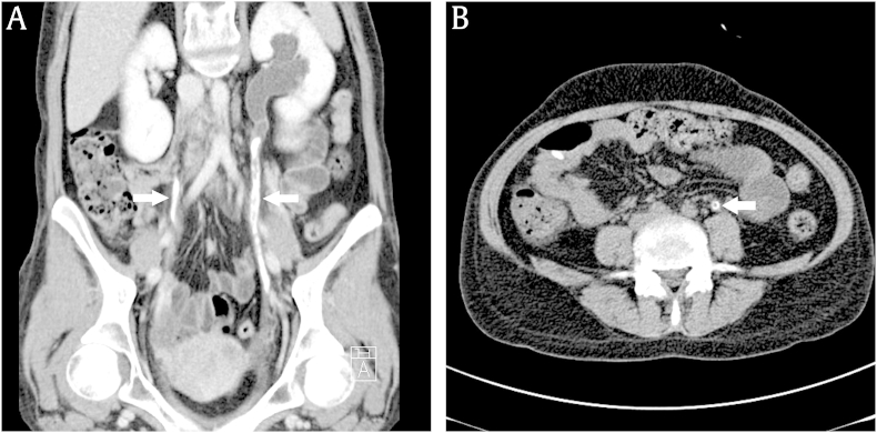
A 23-year-old woman with systemic lupus erythematosus (SLE) was admitted for evaluation of anemia. She was diagnosed with SLE 2┬Āyears earlier after visiting our hospital with swelling and pain in both proximal interphalangeal joints several years in duration. Although symptoms improved while taking prednisolone and hydroxychloroquine, these medications were discontinued after 10 months because of pregnancy. Eight months before admission, she was treated with prednisolone for SLE flare with thrombocytopenia, elevated liver function, and pulmonary congestion after giving birth. After recovery, hydroxychloroquine and mycophenolate mofetil were added, and she has been stable since then. She was hospitalized because of asymptomatic hemoglobin decrease. However, she complained of left flank pain after admission. Physical examination showed no costovertebral angle tenderness. Urinalysis revealed 2+ albumin, many red blood cells per high power field, and many white blood cells per high power field. Laboratory examination revealed hemoglobin 7.3┬Āmg/dL, creatinine 1.3┬Āmg/dL, total calcium 9.0┬Āmg/dL, inorganic phosphate 2.8┬Āmg/dL, parathyroid hormone 77.8┬Āpg/dL, and C-reactive protein 0.04┬Āmg/dL. Computed tomography┬Āof the abdomen showed bilateral ureteral calcification and stones with hydronephrosis of the left kidney (Fig.┬Ā1).
SLE is a chronic systemic inflammatory autoimmune disease affecting various organ systems. Calcinosis occurs in damaged tissues, and calcification has been reported primarily in the musculoskeletal, cerebral, and vascular systems of SLE patients. Bilateral calcification of the ureters is quite rare; however, when flank pain, hematuria, or azotemia develops in SLE patients, ureteral calcification and obstruction should be considered.
 PDF Links
 PubReader
 Full text via DOI
 Download Citation
 Print
Print


")


