Kidney Res Clin Pract > Volume 43(2); 2024 > Article
Cha: Pharmacologic therapeutics in sarcopenia with chronic kidney disease

Abstract

Inflammation, metabolic acidosis, renin-angiotensin system activation, insulin resistance, and impaired perfusion to skeletal muscles, among others, are possible causes of uremic sarcopenia. These conditions induce the activation of the nuclear factor-kappa B and mitogen-activated protein kinase pathways, adenosine triphosphate ubiquitin-proteasome system, and reactive oxygen species system, resulting in protein catabolism. Strategies for the prevention and treatment of sarcopenia in chronic kidney disease (CKD) are aerobic and resistance exercises along with nutritional interventions. Anabolic hormones have shown beneficial effects. Megestrol acetate increased weight, protein catabolic rate, and albumin concentration, and it increased intracellular water component and muscle mass. Vitamin D supplementation showed improvement in physical function, muscle strength, and muscle mass. Correction of metabolic acidosis showed an increase in protein intake, serum albumin levels, body weight, and mid-arm circumference. The kidney-gut-muscle axis indicates that dysbiosis and changes in gut-derived uremic toxins and short-chain fatty acids affect muscle mass, composition, strength, and functional capacity. Biotic supplements, AST-120 administration, hemodiafiltration, and preservation of residual renal function are alleged to reduce uremic toxins, including indoxyl sulfate (IS) and p-cresyl sulfate (PCS). Synbiotics reversed the microbiota change in CKD patients and decreased uremic toxins. AST-120 administration changed the overall gut microbiota composition in CKD. AST-120 prevented IS and PCS tissue accumulation, ameliorated muscle atrophy, improved exercise capacity and mitochondrial biogenesis, restored epithelial tight junction proteins, and reduced plasma endotoxin levels and markers of oxidative stress and inflammation. In a human study, the addition of AST-120 to standard treatment had modest beneficial effects on gait speed change and quality of life.

Introduction

Sarcopenia is characterized by the loss of muscle mass and strength, decreased physical performance, decreased quality of life (QoL), morbidities, and immobility with age, and it is associated with protein-energy wasting in chronic kidney disease (CKD) and end-stage kidney disease (ESKD) patients [1]. There have been several recommendations for sarcopenia criteria in Europe and Asia (Table 1) [2,3]. These recommendations use muscle strength, muscle quantity or quality, and physical performance to diagnose and classify sarcopenia.
Sarcopenia is primarily the age-related progressive loss of muscle mass and strength (muscle atrophy). Uremic sarcopenia means a progressive decrease in muscle mass, strength, and function despite normal skeletal muscle physiology in CKD patients. Uremic sarcopenia is not solely associated with the advanced age. It is mainly associated with the characteristics of uremia, such as metabolic acidosis, chronic inflammation, vitamin D deficiency, insulin resistance, hormonal changes, and gut dysbiosis, which contribute to the development of increased protein catabolism and reduced protein synthesis.
I will deal with the clinical significance, prevalence, risk factors, and strategies to prevent and treat sarcopenia with CKD in this review.

Clinical significance and prevalence of sarcopenia in chronic kidney disease

Sarcopenia is an important issue in dialysis-independent CKD and ESKD patients because it increases the risk of death, cardiovascular complications, the hospitalization rate, and disabilities. Sarcopenia is a strong predictor of mortality in dialysis-independent CKD and hemodialysis (HD) patients (hazard ratio [HR], 2.89; 95% confidence interval [CI], 1.40–5.96; p < 0.004 and HR, 6.99; 95% CI, 1.84–26.58; p = 0.004, respectively) [4,5]. Sarcopenia is a risk factor for cardiovascular events in HD patients (HR, 4.33; 95% CI, 1.51–12.43; p = 0.006) [5]. Sarcopenia is associated with a higher risk of hospitalization in HD patients (relative risk, 2.07; 95% CI, 1.48–2.88; p < 0.001) [6]. Sarcopenic patients exhibited a higher risk of falls than nonsarcopenic patients [7].
Physical inactivity due to sarcopenia results in poor prognosis in CKD patients. Several reports have shown a significant association of physical inactivity with kidney survival and mortality [8,9]. Therefore, maintaining physical performance to prevent sarcopenia is a critical factor in improving the prognosis of CKD patients. The prevalence and incidence of ESKD in Korea are increasing, with >100,000 and 18,642 patients at the end of 2019, respectively [10]. Therefore, the prevention and treatment of sarcopenia in CKD patients are highly important.
Altogether, sarcopenia is a major public health problem and will be even more important in the future given the aging society. The prevalence of sarcopenia in Korean older adults (mean age of 70.3 years) was 32.5% (total), 15.1% (male), and 42.1% (female) based on the Asian Working Group for Sarcopenia (AWGS) 2019 [11].
The prevalence of sarcopenia increases with CKD stage: 17% in stage 3a, 20% in stage 3b, 29% in stage 4, and 38% in stage 5 [12]. A lower lean mass and appendicular skeletal muscle mass index (ASMI) are correlated with glomerular filtration rate (GFR) decline, and a 1 mL/min/1.73 m2 decrease in GFR was associated with a 0.03 ± 0.01 kg/m2 decrease in the ASMI [13].
The prevalence of sarcopenia ranges from 4% to 42% according to the definition used, the population studied, and the stage of CKD [4,1317]. A sarcopenia prevalence of 5.9% to 14% was found in dialysis-independent CKD patients [4,13], and that of peritoneal dialysis (PD) patients was from 4% to 15.5% based on the European Working Group for Sarcopenia criteria [15,16]. It was from 1.9% to 40% based on AWGS cutoffs in PD and HD patients [14,17].
The issue is the application of these criteria in clinical practice and the lack of recommended cutoff points in specific populations, such as CKD and ESKD patients. CKD is a condition associated with muscle loss [1,1820]. There is a possibility of underestimating the clinical significance of sarcopenia in CKD patients [21].

Risk factors of uremic sarcopenia

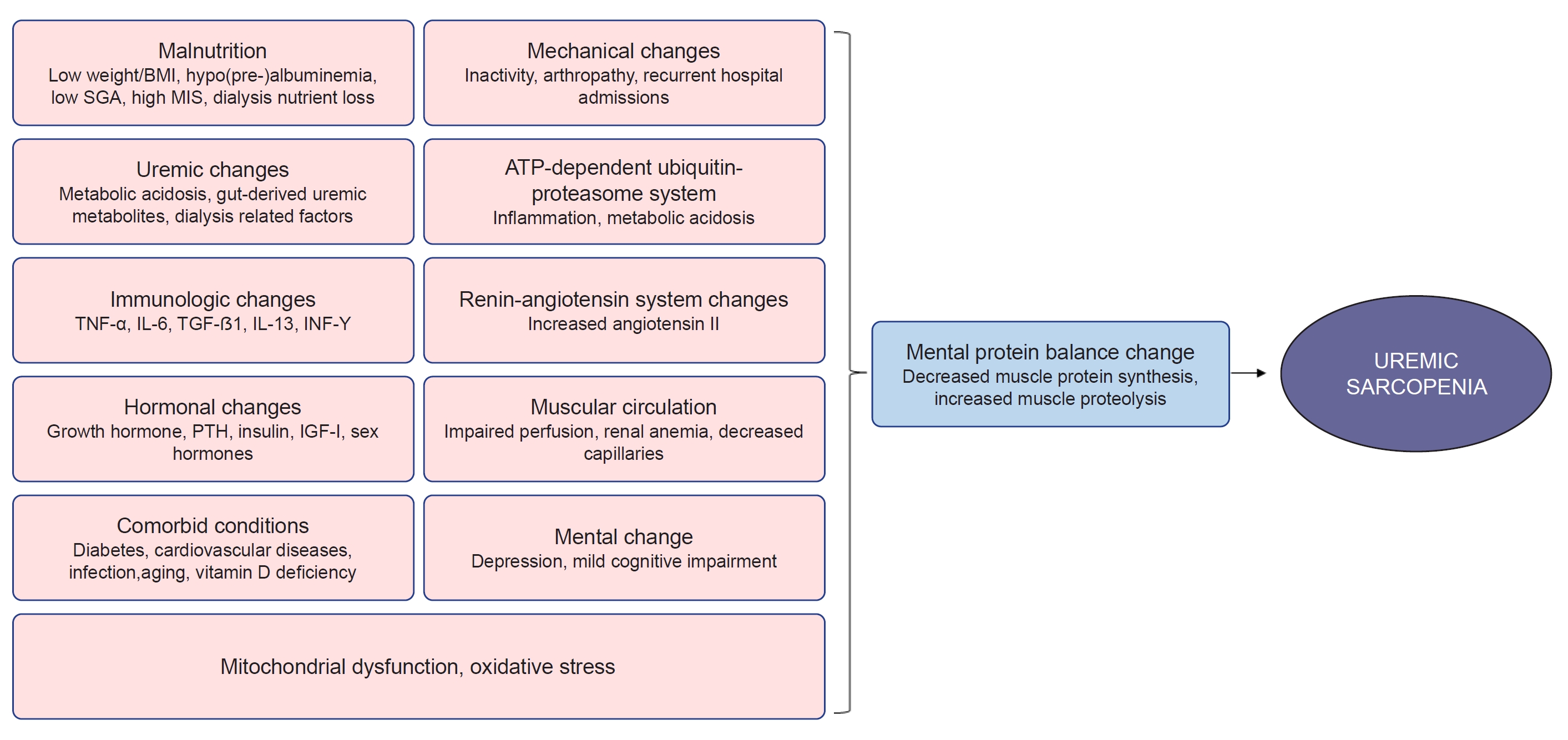
Several risk factors for sarcopenia are alleged in CKD and ESKD patients (Fig. 1).
Aging is associated with sarcopenia and an increased prevalence of CKD, which accelerates normal physiological muscle wasting [22]. CKD is associated with chronic low-grade inflammation leading to progressive weight loss, muscle weakness, and disabilities. Inflammatory cytokines, oxidative stress, and inactivity-mediated destruction of protein homeostasis result in the catabolic destruction of structural and functional proteins, resulting in skeletal muscle wasting and a decrease in exercise capacity [23,24].
Inflammation status, i.e., a high malnutrition-inflammation score (MIS) and high levels of high-sensitive C-reactive protein (CRP), interleukin (IL) 6, β2-microglobulin, and IL-4 is associated with sarcopenia in CKD patients [5,14,2528].
Older age, male sex, low body mass index (BMI), and longer vintage of dialysis are associated with sarcopenia [12,17,29,30]. Malnutrition, i.e., low albumin/prealbumin levels, a high MIS, and poor nutritional status based on subjective global assessment are also associated with sarcopenia [5,14,17,25,27,31,32]. Low serum vitamin D levels and diabetes mellitus are risk factors for sarcopenia [5,12,17,32]. Depression (odds ratio [OR], 6.87; 95% CI, 2.06–22.96; p = 0.002) and mild cognitive impairment (OR, 6.36; 95% CI, 1.62–34.96; p = 0.008) are associated with sarcopenia [25,27].
Impaired perfusion due to chronic heart failure, decreased oxygen supply to skeletal muscles due to anemia, and decreased skeletal muscle capillaries are other risk factors for sarcopenia [33]. Insulin resistance-associated skeletal muscle atrophy and changes in muscle composition, including fat accumulation, are risk factors for sarcopenia in CKD [33,34]. Inactivity and renin-angiotensin system activation are also mechanisms of exercise intolerance in CKD [33].
Mitochondrial dysfunction of skeletal muscle in CKD is considered to be a cause of loss of muscle mass and exercise capacity [3538]. CKD patients have decreased muscle mitochondrial content and oxidative capacity along with suppressed activity of various mitochondrial enzymes, leading to impaired energy production. These changes can be found from the early stages of CKD and are aggravated along with CKD progression [37].
Metabolic acidosis can induce glucocorticoid secretion and insulin resistance and increase branched-chain amino acid oxidation in skeletal muscles, which is associated with decreased muscle protein synthesis [3941]. Acidosis also induces reprogramming of cellular metabolism to mitigate oxidative stress, although this originates from cancer cells [42].

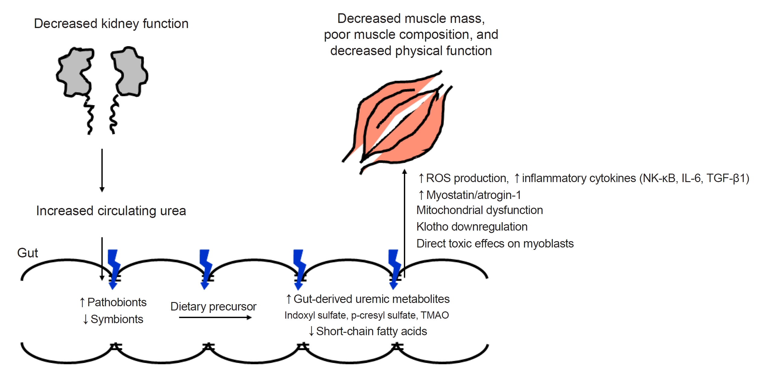
Gut microbiome and uremic metabolite

Influx of urea and other retained toxins exerts a change in the gut microbiome in CKD, favoring pathobiont overgrowth. The abundance of bacteria that contain tryptophanase and urease and produce uremic toxins such as indoxyl sulfate (IS), p-cresyl sulfate (PCS), and trimethylamine-N-oxide (TMAO), increases, while the abundance of beneficial bacteria producing short-chain fatty acids, an essential nutrient for the colonic epithelium, decreases [43]. Lactobacillus and Bifidobacterium are decreased, while Proteobacteria, Enterobacter, Escherichia coli, Acinetobacter, Clostridium perfringens, and Proteus species are increased in the colon of CKD patients [44].
Disruption of the colonic epithelial tight junction associated with urea influx, increased ammonium production, and decreased epithelial survival results in the loss of integrity and increased intestinal permeability, which allows the translocation of bacteria and lipopolysaccharide (LPS). LPS can activate immune cells through the toll-like receptor 4 (TLR-4) dependent and nuclear factor kappa-B (NF-κB) pathway. And pathobionts can stimulate dendritic cells and produce inflammatory cytokines [45]. Prolonged colonic transit time, dietary restriction of fiber, and metabolic acidosis also directly and indirectly contribute to dysbiosis and the altered intestinal environment [46].

Role of the gut microbiome and uremic metabolite on the maintenance of skeletal muscle mass

Reduced muscle mass, altered muscle composition, i.e., increased deposition of lipids or adipocytes within and/or between muscles, and poor physical function are prevalent in ESKD patients. ESKD patients have elevated uremic metabolite levels combined with an altered gut microbiome, increased gut-derived uremic metabolites, and increased epithelial permeability. In turn, the fecal levels of short-chain fatty acids are reduced. The accumulation of gut-derived uremic metabolites within skeletal muscle results in decreased muscle mass or increased lipid content [47]. Increased urea due to decreased kidney function, gut dysbiosis, increased circulating uremic metabolites, and the resultant reduced muscle mass, altered muscle composition, and poor physical function is called the kidney-gut-muscle axis (Fig. 2).
Protein-derived tryptophan is metabolized into indole, which is then absorbed into the bloodstream and oxidized into IS in the liver [48]. Uremic toxins enter target cells via specific transporters, such as the organic anion transporter (OAT), and then, exert their toxicity via the activation of cellular nicotinamide adenine dinucleotide phosphate hydrogen (NADPH) oxidase, which results in the overproduction of reactive oxygen species (ROS) and inflammatory cytokines [49,50]. IS also enters various cells via OATs (OAT1 and OAT3), and OATs are expressed in muscles [5153].
IS and PCS accumulate in various organs, including skeletal muscle, in the CKD mouse model [54]. IS exposure inhibited cell proliferation and cell viability and decreased mitochondrial function in mouse muscle cells. CKD patients also showed a significant inverse association between plasma IS levels and skeletal muscle mass [55]. High IS levels were associated with longer HD vintage, and IS was negatively correlated with the skeletal muscle mass index in HD patients. HD patients with high IS levels showed greater loss of hand grip strength (HGS) [56].
Accumulated IS in muscle cells via OAT activates NADPH oxidase and the aryl hydrocarbon receptor (AHR) pathway to cause increased ROS production. Enhanced ROS trigger inflammatory cytokines, such as tumor necrosis factor alpha (TNF-α), IL-6, and transforming growth factor beta 1 (TGF-β1), and induce myostatin (negative regulator of muscle growth) and atrogin-1 (muscle-specific ubiquitin ligase family) expression, which are involved in muscle wasting [52].
IS also induces mitochondrial network disintegration through metabolic alterations, such as an increase in antioxidative responses (pentose phosphate pathway and glutathione metabolism), and a decrease in energy generation-related pathways (tricarboxylic acid cycle, glutamine metabolism, and mitochondrial oxidative phosphorylation) in muscle cells, which results in reduced adenosine triphosphate (ATP) production [55]. Muscle mitochondrial dysfunction was found from the early stage of CKD and muscle atrophy followed by exercise impairment [37]. Muscular mitochondria were decreased in the early stage of CKD in mice.
In addition, IS downregulated Klotho expression through ROS-associated NF-κB activation [57]. Klotho levels and skeletal muscle physiology are closely related [58,59]. Klotho-deficient mice showed significantly decreased body weight and forelimb grip strength, and Klotho overexpression restored grip strength and running capacity [60]. It is possible that downregulation of Klotho by IS in muscles is one of the mechanisms of sarcopenia in CKD. IS also has direct toxic effects on myoblasts by decreasing their viability and increasing cell apoptosis [61].
A systematic review and meta-analysis showed that biotic supplements, AST-120, hemodiafiltration, and preservation of residual renal function significantly reduced uremic toxins, including IS and PCS [62]. A reduction in the elevated AHR transactivating activity by dialysis was also reported [63].

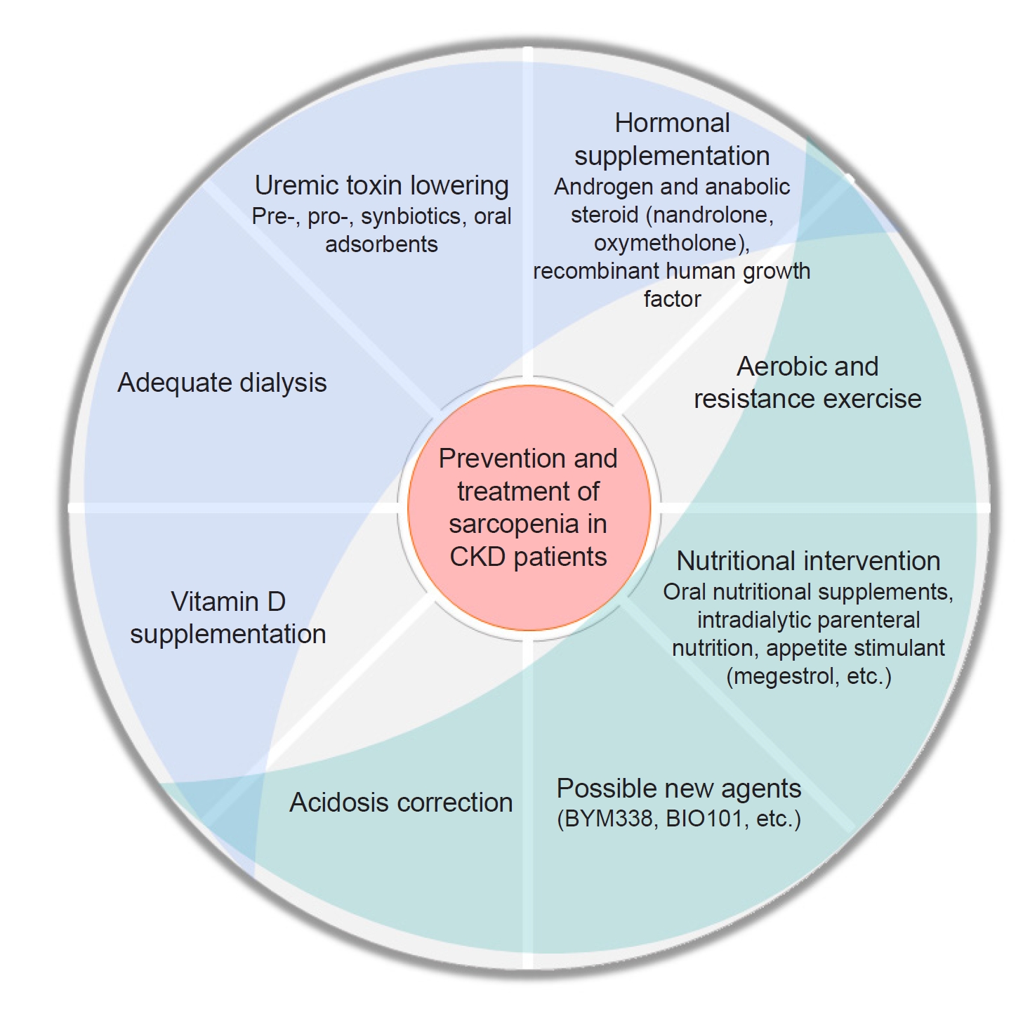
Strategies to prevent and treat sarcopenia

Researchers can acquire an impression regarding the treatment strategies based on all of the risk factors mentioned above. Many drugs have been developed to prevent or treat sarcopenia. However, there are no drugs approved by the U.S. Food and Drug Administration for the treatment of sarcopenia [64]. Only a few of them, i.e., Bimagrumab from Novartis and Sarconeos from Biophytis SAS, have recently shown favorable results.
Bimagrumab (BYM338) is a fully human monoclonal antibody, which was developed to treat pathological muscle loss and weakness. It binds to and inhibits activin receptor type-2B and prevents the actions of natural ligands that negatively regulate skeletal muscle growth [65,66]. Activin receptor type-2B blockade with Bimagrumab resulted in the significant loss of fat mass, gain in lean mass, and metabolic improvements during 48 weeks in patients with overweight or obesity who had type 2 diabetes in a phase 2 trial [66].
20‐Hydroxyecdysone (20E) is a polyhydroxylated plant steroid that has pharmacological effects in aging and sarcopenia. Sarconeos (BIO101) is a 20E purified investigational drug (≥97%). It targets proto-oncogene protein-c-MAS-1, MAS receptor. BIO101 showed a good safety and pharmacokinetic profile in a phase 1 study for healthy young and older adults [67]. In a phase 2 study (NCT03452488), BIO101 was orally administered for 26 weeks to community-dwelling men and women aged ≥65 years, suffering from age-related sarcopenia (including sarcopenic obesity), and at risk of mobility disability. It was a double-blind, placebo-controlled clinical trial. BIO101 at the highest dose (350 mg, twice daily) showed a clinically meaningful improvement in the 400-m walk test [68].
The main interventions for sarcopenia in CKD patients are aerobic and resistance exercises along with nutritional interventions. Furthermore, optimizing dialysis, vitamin D status, acidosis, and the management of comorbidities such as depression are mandatory [69].

Megestrol acetate

HD patients with no residual kidney function (age of 40–80 years) treated with a daily dose of 160 mg of megestrol acetate for 2 to 12 months, experienced an increase in weight (2–9 kg) associated with an increase in the protein catabolic rate, albumin concentration, serum creatinine and urea [70]. In addition, megestrol improved body water distribution with an increase in the intracellular water component and muscle mass.
A review of small retrospective studies showed an increase in BMI, albumin levels, and protein-energy intake after megestrol acetate [71]. A systematic review, especially of dialysis patients, showed a significant increase in body weight, albumin levels, and appetite, although they were small studies (n = 9–32) with a short duration (8–24 weeks) and a high degree of bias. Megestrol acetate was also associated with overhydration, excessive fluid gain, diarrhea, hyperglycemia, suppressed cortisol levels, thrombophlebitis, nausea, and vomiting [72].

Hormonal therapies

Nandrolone (decanoate), an androgen and anabolic steroid medication, which is used in the treatment of wasting syndrome and is given through intramuscular or subcutaneous way, injection in HD patients for 6 months significantly increased lean body mass, improved functional capacity (walking and stair-climbing test), and increased appendicular lean mass in a dose-responsive manner [73,74]. Nandrolone combined with intradialytic resistance training for 12 weeks increased the quadriceps muscle cross-sectional area in an additive manner [75].
Oxymetholone, an oral androgen and anabolic steroid, which is also used in the treatment of wasting syndrome and promotion of weight gain and muscle growth, has a higher anabolic effect and lower androgenic activity than testosterone. HD patients showed an increase in fat-free mass, HGS, and physical functioning scores as well as an increase in mRNA expression for myosin heavy chain, and insulin-like growth factor (IGF)-I/II in muscles after oxymetholone administration for 24 weeks [76].
Recombinant human growth hormone (rhGH) increased protein synthesis and decreased protein catabolism in HD and PD patients [7780]. rhGH increased lean body mass and improved QoL [81]. rhGH also increased HGS after 6 months in elderly HD patients [82]. However, another large randomized controlled trial (RCT) study (OPPORTUNITY) failed to show an increase in albumin levels, lean body mass, physical capacity, or QoL [83]. rhGH also showed adverse effects, including soft tissue edema, arthralgia, carpal tunnel syndrome, gynecomastia, and dysglycemia [83].

Vitamin D

CKD patients in stages 3 to 4 and PD patients with vitamin D deficiency became vitamin D sufficient after vitamin D replacement. They showed improvements in physical function (timed up and go, gait speed, timed chair stand, stair climb, static and dynamic balance) and isometric strength [84]. Vitamin D supplementation in male HD patients also improved muscle mass [85].
However, supplementation of cholecalciferol 50,000 units/week with the target of 25(OH)D concentration >80 nmol/L in HD patients with an initial 25(OH)D concentration <50 nmol/L had no effect on muscle strength or symptoms [86]. Treatment with 50,000 units of oral cholecalciferol supplementation, once weekly for 8 weeks and then monthly for 4 months, to HD patients with a 25(OH)D concentration <60 nmol/L showed higher 25(OH)D and 1,25(OH)2D levels without increased calcium or phosphorus levels. However, muscle strength, functional capacity, and QoL did not change [87].

Correction of acidosis

Advanced CKD patients (eGFR of 15–30 mL/min/1.73 m2) with metabolic acidosis presented an increase in the mid-arm circumference, protein intake, and serum albumin levels after oral sodium bicarbonate administration for 2 years [88]. An RCT of PD patients showed an increased body weight and mid-arm circumference and decreased hospitalization after 1 year of high alkali dialysate [89]. In addition, a high bicarbonate dialysate concentration of 40 mmol/L resulted in better control of acidosis and increased triceps skin fold thickness compared to a bicarbonate concentration of 30 mmol/L [90].

Biotics

Because gut dysbiosis is associated with systemic inflammation, the administration of biotics can be an indirect target to ameliorate muscle wasting. Lactobacillus species and Bifidobacterium ameliorated muscle wasting in mouse models, and Lactobacillus exhibited the potential to restore microbiome balance, and the gut permeability, which was associated with the improvement in muscle mass or strength, was reduced [91]. However, probiotics containing Lactobacillus and Bifidobacterium did not show meaningful effects on muscle mass and function in humans, because of the scarcity of studies, the variability in the populations, and the difficulty in accurately and reproducibly measuring muscle mass and function [92].
Nonetheless, Bifidobacterium longum was significantly increased after prebiotic administration in super-elderly patients with sarcopenia. The skeletal muscle mass index was increased, while the body fat percentage was decreased [93]. A randomized, double-blind, placebo-controlled, crossover trial using synbiotics over 6 weeks in predialysis CKD patients showed an increase in Bifidobacterium and Lactobacillus after synbiotic therapy as well as a decrease in Clostridiales. The Bifidobacterium change showed an inverse correlation with the concentration of serum IS and PCS [94].
Although there is no data about the effect of biotics on muscle mass, muscle strength, or physical function, there are several reports about the relationship between biotics and surrogate outcomes such as inflammation, oxidative stress, uremic toxins, and endotoxins.
Probiotic supplementation in HD patients reduced systemic inflammatory responses, which was associated with an increase in Tregs and a decrease in proinflammatory monocytes [95]. Probiotic, prebiotic, and synbiotic administration also reduced CRP, IL-6, and IS levels, and increased high-density lipoprotein cholesterol in dialysis patients [96]. Synbiotics were more effective than probiotics in improving inflammatory markers, endotoxins, and anti-heat shock protein70 serum levels in HD patients [97]. Synbiotics also reduced serum IS, PCS, and urea in HD patients [98].

Oral adsorbents

AST-120 adsorbs uremic toxins and precursors, including indole, and excretes them into the feces.
AST-120 administration increased Lactobacillus in CKD rats. The microbiota composition after AST-120 treatment was between that of normal and CKD rats [99]. CKD rats showed elevated plasma endotoxin, IL-6, TNF-α, monocyte chemoattractant protein-1 (MCP-1), cytokine-induced neutrophil chemoattractant-3 (CINC-3), L-selectin, intercellular adhesion molecule-1 (ICAM-1), and malondialdehyde levels and depletion of the colonic epithelial tight junction proteins, claudin-1, occludin, and zonular occludens-1 (ZO-1). AST-120 showed partial restoration of epithelial tight junction proteins and a reduction in plasma endotoxin and markers of oxidative stress and inflammation [100].
AST-120 prevented the tissue accumulation of IS and PCS in skeletal muscle, which resulted in the amelioration of muscle atrophy (cross-sectional area) [54]. AST-120 prevented CKD-induced physical inactivity mainly by maintaining mitochondrial function, suppressing atrogin-1/myostatin expression, and recovering Akt phosphorylation in skeletal muscle [101]. A reduction in oxidative stress via AST-120 improved exercise capacity and mitochondrial biogenesis of skeletal muscle [33,101]. AST-120 significantly restored skeletal muscle weight in CKD mice. In addition, AST-120 increased the renal expression of Klotho, which was decreased under uremic conditions [102]. Klotho levels and skeletal muscle physiology were closely related, as we mentioned above [58,59].
In a human RCT, gait speed significantly increased over 1 year after carbonaceous oral adsorbent administration. Bodily pain, vitality, symptoms/problems, and cognitive function also improved in the oral adsorbent group, while the quality of social interactions and kidney disease effects decreased in the control group [103]. Prospective studies with longer follow-up durations, larger sample sizes, and interventions to lower uremic toxins are necessary to elucidate the role of uremic toxins in CKD patients with sarcopenia.
The effectiveness of pharmacologic interventions mentioned above is summarized in Table 2.
In conclusion, aerobic and resistance exercise, along with nutritional interventions, including oral and intradialytic parenteral nutritional supplements, has been the main preventive or treatment strategies for sarcopenia in dialysis-independent CKD and ESKD patients. In addition, anabolic hormones, including anabolic steroids and growth hormones, and appetite stimulants showed beneficial effects on biochemical markers such as serum albumin level, both muscle strength and mass, and physical function. Correction of metabolic acidosis and vitamin D status also showed favorable effects on physical function, muscle mass, and strength. Biotics improved the gut microbiome composition and the level of uremic toxins, endotoxins, inflammation, and oxidative stress. Oral adsorbents also showed beneficial effects on the restoration of the gut microbiome composition and intestinal wall integrity as well as on the skeletal muscle physiology. A multifaceted approach can be helpful to lessen the burden of sarcopenia in CKD patients (Fig. 3).

Notes

Conflicts of interest

The author has no conflicts of interest to declare.

Data sharing statement

The data presented in this study are available on request from the corresponding author.

Figure 1.

Possible etiologies of uremic sarcopenia.

ATP, adenosine triphosphate; BMI, body mass index; IGF, insulin-like growth factor; IL-6, interleukin 6; IL-1β, interleukin-1β; INF-γ, interferon-γ; MIS, malnutrition-inflammation score; PTH, parathyroid hormone; SGA, subjective global assessment; TGF-β1, transforming growth factor beta 1; TNF-α, tumor necrosis factor alpha.
j-krcp-23-094f1.jpg
Figure 2.

Scheme of the kidney-gut-muscle axis.

IL-6, interleukin 6; NF-κB, nuclear factor kappa-B; ROS, reactive oxygen species; TGF-β1, transforming growth factor beta 1; TMAO, trimethylamine-N-oxide.
j-krcp-23-094f2.jpg
Figure 3.

Multifaceted approach for sarcopenia in CKD patients.

CKD, chronic kidney disease.
j-krcp-23-094f3.jpg
Table 1.
European and Asian criteria for sarcopenia
European Working Group on Sarcopenia in older people 2 (EWGSOP2) Asian Working Group for Sarcopenia (AWGS) 2019
(Case finding) Clinical suspicion or SARC-F (Case finding) Clinical conditions: functional decline or limitation, unintentional weight loss, depressive mood, cognitive impairment, repeated falls, malnutrition
Chronic conditions: heart failure, chronic obstructive pulmonary disease, diabetes mellitus, chronic kidney disease, etc.
No clinical conditions: calf circumference (male, <34 cm; female, <33 cm) or SARC-F ≥4 or SARC-CalF ≥11
Cutoff for men Cutoff for women Cutoff for men Cutoff for women
(Assess) (Diagnosis)
 HGS <27 kg <16 kg  HGS <28 kg <18 kg
 Chair stand >15 sec/5 rises  Physical performance
(Confirm)   6-m walk speed <1.0 m/sec
 ASM <20 kg <15 kg   Chair stand ≥12 sec/5 rises
 ASM/height2 <7.0 kg/m2 <5.5 kg/m2   SPPB ≤9 point score
(Severity)  Muscle quantity
 4-m gait speed ≤0.8 m/sec   DEXA <7.0 kg/m2 <5.4 kg/m2
 SPPB ≤8 point score   BIA <7.0 kg/m2 <5.7 kg/m2
 TUG ≥20 sec (Severity)
 400-m walk test Non-completion or ≥6 min for completion  Sarcopenia Low ASM + low muscle strength or low physical performance
 Severe sarcopenia Low ASM + low muscle strength and low physical performance

ASM, appendicular skeletal muscle mass; BIA, bioimpedance analysis; DEXA, dual-energy X-ray absorptiometry; HGS, handgrip strength; SARC-F, screening tool for sarcopenia, questionnaire consisting of 5 questions: strength (S), assistance walking (A), rising from a chair (R), climbing stairs (C), and falls (F) on a scale of 0 to 2; SARC-CalF, SARC-F + calf circumference; SPPB, short physical performance battery; TUG, timed up and go.

Table 2.
Effectiveness of pharmacologic interventions for sarcopenia, especially with CKD
Drug Patient Result
Megestrol acetate [4951] HD patients ↑Appetite, ↑weight/BMI, ↑protein catabolic rate, ↑serum albumin/creatinine/urea
↑Intracellular water component
↑Muscle mass
Nandrolone [5254] HD patients ↑Lean body mass
↑Walking speed, stair-climbing
↑Appendicular lean mass
↑Quadriceps muscle cross-sectional area (+resistance training)
Oxymetholone [55] HD patients ↑Fat-free mass, HGS, physical functioning scores
↑mRNA expression for myosin heavy chain
↑IGF-I/II
Recombinant human growth factor [5660] HD and PD patients ↑Protein synthesis, ↓protein catabolism
↑Lean body mass, HGS
↑QoL
Vitamin D [63, 64] CKD stage 3–4 and PD patients with vitamin D deficiency ↑Vitamin D concentration
HD patients ↑Physical functioning (TUG, gait speed, timed chair stand, stair climbing, static/dynamic balance)
↑Isometric strength, muscle mass
Bicarbonate [6769] Glomerular filtration rate 15–30 mL/min/1.73 m2 with metabolic acidosis ↑Mid-arm circumference, protein intake, serum albumin
PD patients ↑Weight, mid-arm circumference
HD patients ↓Hospitalization
↑Acidosis control, triceps skin fold thickness
Prebiotics [93] Super-elderly with sarcopenia Bifidobacterium longum
↑Skeletal muscle index, ↓body fat percentage
Synbiotics [94] Predialysis CKD patients Bifidobacterium, Lactobacillus, ↓Clostridium
↓Serum indoxyl sulfate, p-cresyl sulfate
Probiotics [95] HD patients ↓Systemic inflammation
Pre-, pro-, synbiotics [96] Dialysis patients ↓C-reactive protein, interleukin-6, indoxyl sulfate, ↑high-density lipoprotein
Synbiotics [97,98] HD patients ↓Inflammation marker, endotoxin, anti-heat shock protein70
↓Indoxyl sulfate, p-cresyl sulfate, urea
AST-120 [103] Advanced CKD patients with serum creatinine between 2.0 and 5.0 mg/dL ↑Gait speed
↑Bodily pain, vitality, symptoms/problems, cognitive function scores
Bimagrumab (BYM338) [45] Overweight or obese type 2 diabetes ↓Fat mass, ↑lean mass
↓HbA1c, ↑QUICK
→HOMA2 (36-week), →Matsuda index (48-week)
Sarconeos (BIO101) [47] Community-dwelling men and women aged ≥65 years, age-related sarcopenia, at risk of mobility disability ↑400-m walk test

BMI, body mass index; CKD, chronic kidney disease; HbA1c, hemoglobin A1c; HD, hemodialysis; HGS, handgrip strength; HOMA, homeostatic model assessment; IGF, insulin-like growth factor; mRNA, messenger RNA; PD, peritoneal dialysis; QoL, quality of life; QUICK, quantitative insulin sensitivity check index; TUG, timed up and go.

Bimagrumab, Novartis; Sarconeos, Biophytis SAS.

References

1. Moon SJ, Kim TH, Yoon SY, Chung JH, Hwang HJ. Relationship between stage of chronic kidney disease and sarcopenia in Korean aged 40 years and older using the Korea National Health and Nutrition Examination Surveys (KNHANES IV-2, 3, and V-1, 2), 2008-2011. PLoS One 2015;10:e0130740.
crossref pmid pmc
2. Cruz-Jentoft AJ, Bahat G, Bauer J, et al. Sarcopenia: revised European consensus on definition and diagnosis. Age Ageing 2019;48:16–31.
crossref pmid pdf
3. Chen LK, Woo J, Assantachai P, et al. Asian Working Group for Sarcopenia: 2019 consensus update on sarcopenia diagnosis and treatment. J Am Med Dir Assoc 2020;21:300–307.
crossref pmid
4. Pereira RA, Cordeiro AC, Avesani CM, et al. Sarcopenia in chronic kidney disease on conservative therapy: prevalence and association with mortality. Nephrol Dial Transplant 2015;30:1718–1725.
crossref pmid
5. Kim JK, Kim SG, Oh JE, et al. Impact of sarcopenia on long-term mortality and cardiovascular events in patients undergoing hemodialysis. Korean J Intern Med 2019;34:599–607.
crossref pmid pdf
6. Giglio J, Kamimura MA, Lamarca F, Rodrigues J, Santin F, Avesani CM. Association of sarcopenia with nutritional parameters, quality of life, hospitalization, and mortality rates of elderly patients on hemodialysis. J Ren Nutr 2018;28:197–207.
crossref pmid
7. Aly SH, Tawfik HM, Bichari WA, Alsadany MA, Banouby E, Hassan M. Sarcopenia and risk of falls in patients with chronic kidney disease, a meet of three geriatric giants. Egypt J Geriatr Gerontol 2019;6:1–5.
crossref
8. Carrero JJ, Chmielewski M, Axelsson J, et al. Muscle atrophy, inflammation and clinical outcome in incident and prevalent dialysis patients. Clin Nutr 2008;27:557–564.
crossref pmid
9. Beddhu S, Wei G, Marcus RL, Chonchol M, Greene T. Light-intensity physical activities and mortality in the United States general population and CKD subpopulation. Clin J Am Soc Nephrol 2015;10:1145–1153.
crossref pmid pmc
10. Hong YA, Ban TH, Kang CY, et al. Trends in epidemiologic characteristics of end-stage renal disease from 2019 Korean Renal Data System (KORDS). Kidney Res Clin Pract 2021;40:52–61.
crossref pmid pmc pdf
11. Seo AR, Kim MJ, Kim B, et al. Associations between frailty in older adults and malnutrition in rural areas: 2019 updated version of the Asian Working Group for Sarcopenia. Yonsei Med J 2021;62:249–254.
crossref pmid pmc pdf
12. Ishikawa S, Naito S, Iimori S, et al. Loop diuretics are associated with greater risk of sarcopenia in patients with non-dialysis-dependent chronic kidney disease. PLoS One 2018;13:e0192990.
crossref pmid pmc
13. Zhou Y, Hellberg M, Svensson P, Höglund P, Clyne N. Sarcopenia and relationships between muscle mass, measured glomerular filtration rate and physical function in patients with chronic kidney disease stages 3-5. Nephrol Dial Transplant 2018;33:342–348.
crossref pmid
14. Kamijo Y, Kanda E, Ishibashi Y, Yoshida M. Sarcopenia and frailty in PD: impact on mortality, malnutrition, and inflammation. Perit Dial Int 2018;38:447–454.
crossref pmid pdf
15. Abro A, Delicata LA, Vongsanim S, Davenport A. Differences in the prevalence of sarcopenia in peritoneal dialysis patients using hand grip strength and appendicular lean mass: depends upon guideline definitions. Eur J Clin Nutr 2018;72:993–999.
crossref pmid pdf
16. da Silva MZ, Vogt BP, Reis NS, Caramori JC. Update of the European consensus on sarcopenia: what has changed in diagnosis and prevalence in peritoneal dialysis? Eur J Clin Nutr 2019;73:1209–1211.
crossref pmid pdf
17. Mori K, Nishide K, Okuno S, et al. Impact of diabetes on sarcopenia and mortality in patients undergoing hemodialysis. BMC Nephrol 2019;20:105.
crossref pmid pmc pdf
18. Fried LF, Boudreau R, Lee JS, et al. Kidney function as a predictor of loss of lean mass in older adults: health, aging and body composition study. J Am Geriatr Soc 2007;55:1578–1584.
crossref pmid
19. Kim H, Suzuki T, Kim M, et al. Incidence and predictors of sarcopenia onset in community-dwelling elderly Japanese women: 4-year follow-up study. J Am Med Dir Assoc 2015;16:85.
crossref
20. Liu CK, Lyass A, Massaro JM, D’Agostino RB Sr, Fox CS, Murabito JM. Chronic kidney disease defined by cystatin C predicts mobility disability and changes in gait speed: the Framingham Offspring Study. J Gerontol A Biol Sci Med Sci 2014;69:301–307.
crossref pmid
21. Studenski SA, Peters KW, Alley DE, et al. The FNIH sarcopenia project: rationale, study description, conference recommendations, and final estimates. J Gerontol A Biol Sci Med Sci 2014;69:547–558.
crossref pmid pmc
22. Fahal IH. Uraemic sarcopenia: aetiology and implications. Nephrol Dial Transplant 2014;29:1655–1665.
crossref pmid
23. Lenk K, Schuler G, Adams V. Skeletal muscle wasting in cachexia and sarcopenia: molecular pathophysiology and impact of exercise training. J Cachexia Sarcopenia Muscle 2010;1:9–21.
crossref pmid pmc
24. Wang XH, Mitch WE. Mechanisms of muscle wasting in chronic kidney disease. Nat Rev Nephrol 2014;10:504–516.
crossref pmid pmc pdf
25. Vettoretti S, Caldiroli L, Armelloni S, Ferrari C, Cesari M, Messa P. Sarcopenia is associated with malnutrition but not with systemic inflammation in older persons with advanced CKD. Nutrients 2019;11:1378.
crossref pmid pmc
26. Souza VA, Oliveira D, Barbosa SR, et al. Sarcopenia in patients with chronic kidney disease not yet on dialysis: analysis of the prevalence and associated factors. PLoS One 2017;12:e0176230.
crossref pmid pmc
27. Kim JK, Choi SR, Choi MJ, et al. Prevalence of and factors associated with sarcopenia in elderly patients with end-stage renal disease. Clin Nutr 2014;33:64–68.
crossref pmid
28. Tabibi H, As’habi A, Najafi I, Hedayati M. Prevalence of dynapenic obesity and sarcopenic obesity and their associations with cardiovascular disease risk factors in peritoneal dialysis patients. Kidney Res Clin Pract 2018;37:404–413.
crossref pmid pmc
29. Bataille S, Serveaux M, Carreno E, Pedinielli N, Darmon P, Robert A. The diagnosis of sarcopenia is mainly driven by muscle mass in hemodialysis patients. Clin Nutr 2017;36:1654–1660.
crossref pmid
30. As’habi A, Najafi I, Tabibi H, Hedayati M. Prevalence of sarcopenia and dynapenia and their determinants in iranian peritoneal dialysis patients. Iran J Kidney Dis 2018;12:53–60.
pmid
31. Isoyama N, Qureshi AR, Avesani CM, et al. Comparative associations of muscle mass and muscle strength with mortality in dialysis patients. Clin J Am Soc Nephrol 2014;9:1720–1728.
crossref pmid pmc
32. Ren H, Gong D, Jia F, Xu B, Liu Z. Sarcopenia in patients undergoing maintenance hemodialysis: incidence rate, risk factors and its effect on survival risk. Ren Fail 2016;38:364–371.
crossref pmid
33. Nishikawa M, Ishimori N, Takada S, et al. AST-120 ameliorates lowered exercise capacity and mitochondrial biogenesis in the skeletal muscle from mice with chronic kidney disease via reducing oxidative stress. Nephrol Dial Transplant 2015;30:934–942.
crossref pmid
34. Park HS, Lim JS, Lim SK. Determinants of bone mass and insulin resistance in Korean postmenopausal women: muscle area, strength, or composition? Yonsei Med J 2019;60:742–750.
crossref pmid pmc pdf
35. Adey D, Kumar R, McCarthy JT, Nair KS. Reduced synthesis of muscle proteins in chronic renal failure. Am J Physiol Endocrinol Metab 2000;278:E219–E225.
crossref pmid
36. Yazdi PG, Moradi H, Yang JY, Wang PH, Vaziri ND. Skeletal muscle mitochondrial depletion and dysfunction in chronic kidney disease. Int J Clin Exp Med 2013;6:532–539.
pmid pmc
37. Tamaki M, Miyashita K, Wakino S, Mitsuishi M, Hayashi K, Itoh H. Chronic kidney disease reduces muscle mitochondria and exercise endurance and its exacerbation by dietary protein through inactivation of pyruvate dehydrogenase. Kidney Int 2014;85:1330–1339.
crossref pmid
38. Ertuglu L, Yildiz A, Gamboa J, Ikizler TA. Skeletal muscle energetics in patients with moderate to advanced kidney disease. Kidney Res Clin Pract 2022;41:14–21.
crossref pmid pmc pdf
39. Kalantar-Zadeh K, Mehrotra R, Fouque D, Kopple JD. Metabolic acidosis and malnutrition-inflammation complex syndrome in chronic renal failure. Semin Dial 2004;17:455–465.
pmid
40. Kalantar-Zadeh K, Fouque D. Nutritional management of chronic kidney disease. N Engl J Med 2017;377:1765–1776.
crossref pmid
41. Bailey JL, Zheng B, Hu Z, Price SR, Mitch WE. Chronic kidney disease causes defects in signaling through the insulin receptor substrate/phosphatidylinositol 3-kinase/Akt pathway: implications for muscle atrophy. J Am Soc Nephrol 2006;17:1388–1394.
pmid
42. Lamonte G, Tang X, Chen JL, et al. Acidosis induces reprogramming of cellular metabolism to mitigate oxidative stress. Cancer Metab 2013;1:23.
crossref pmid pmc pdf
43. Lau WL, Savoj J, Nakata MB, Vaziri ND. Altered microbiome in chronic kidney disease: systemic effects of gut-derived uremic toxins. Clin Sci (Lond) 2018;132:509–522.
crossref pmid pdf
44. Cigarran Guldris S, González Parra E, Cases Amenós A. Gut microbiota in chronic kidney disease. Nefrologia 2017;37:9–19.
crossref pmid
45. Al Khodor S, Shatat IF. Gut microbiome and kidney disease: a bidirectional relationship. Pediatr Nephrol 2017;32:921–931.
crossref pmid pdf
46. Kim MG, Yang J, Jo SK. Intestinal microbiota and kidney diseases. Kidney Res Clin Pract 2021;40:335–343.
crossref pmid pmc pdf
47. Lustgarten MS. The kidney-gut-muscle axis in end-stage renal disease is similarly represented in older adults. Nutrients 2019;12:106.
crossref pmid pmc
48. Niwa T, Yazawa T, Maeda K, et al. Effect of oral sorbent, AST-120, on serum concentration of indoxyl sulfate in uremic rats. Nihon Jinzo Gakkai Shi 1990;32:695–701.
pmid
49. Niwa T. Role of indoxyl sulfate in the progression of chronic kidney disease and cardiovascular disease: experimental and clinical effects of oral sorbent AST-120. Ther Apher Dial 2011;15:120–124.
crossref pmid
50. Watanabe H, Miyamoto Y, Honda D, et al. p-Cresyl sulfate causes renal tubular cell damage by inducing oxidative stress by activation of NADPH oxidase. Kidney Int 2013;83:582–592.
crossref pmid
51. Deguchi T, Ohtsuki S, Otagiri M, et al. Major role of organic anion transporter 3 in the transport of indoxyl sulfate in the kidney. Kidney Int 2002;61:1760–1768.
crossref pmid
52. Enoki Y, Watanabe H, Arake R, et al. Indoxyl sulfate potentiates skeletal muscle atrophy by inducing the oxidative stress-mediated expression of myostatin and atrogin-1. Sci Rep 2016;6:32084.
crossref pmid pmc pdf
53. Motojima M, Hosokawa A, Yamato H, Muraki T, Yoshioka T. Uremic toxins of organic anions up-regulate PAI-1 expression by induction of NF-kappaB and free radical in proximal tubular cells. Kidney Int 2003;63:1671–1680.
pmid
54. Sato E, Saigusa D, Mishima E, et al. Impact of the oral adsorbent AST-120 on organ-specific accumulation of uremic toxins: LC-MS/MS and MS imaging techniques. Toxins (Basel) 2017;10:19.
crossref pmid pmc
55. Sato E, Mori T, Mishima E, et al. Metabolic alterations by indoxyl sulfate in skeletal muscle induce uremic sarcopenia in chronic kidney disease. Sci Rep 2016;6:36618.
crossref pmid pmc pdf
56. Lin YL, Liu CH, Lai YH, et al. Association of serum indoxyl sulfate levels with skeletal muscle mass and strength in chronic hemodialysis patients: a 2-year longitudinal analysis. Calcif Tissue Int 2020;107:257–265.
crossref pmid pdf
57. Shimizu H, Bolati D, Adijiang A, et al. Indoxyl sulfate downregulates renal expression of Klotho through production of ROS and activation of nuclear factor-ĸB. Am J Nephrol 2011;33:319–324.
crossref pmid pdf
58. Avin KG, Coen PM, Huang W, et al. Skeletal muscle as a regulator of the longevity protein, Klotho. Front Physiol 2014;5:189.
crossref pmid pmc
59. Semba RD, Ferrucci L, Sun K, et al. Low plasma klotho concentrations and decline of knee strength in older adults. J Gerontol A Biol Sci Med Sci 2016;71:103–108.
crossref pmid
60. Phelps M, Pettan-Brewer C, Ladiges W, Yablonka-Reuveni Z. Decline in muscle strength and running endurance in klotho deficient C57BL/6 mice. Biogerontology 2013;14:729–739.
crossref pmid pdf
61. Rodrigues GG, Dellê H, Brito RB, et al. Indoxyl sulfate contributes to uremic sarcopenia by inducing apoptosis in myoblasts. Arch Med Res 2020;51:21–29.
crossref pmid
62. Takkavatakarn K, Wuttiputinun T, Phannajit J, Praditpornsilpa K, Eiam-Ong S, Susantitaphong P. Protein-bound uremic toxin lowering strategies in chronic kidney disease: a systematic review and meta-analysis. J Nephrol 2021;34:1805–1817.
crossref pmid pdf
63. Kim JT, Kim SH, Min HK, et al. Effect of dialysis on aryl hydrocarbon receptor transactivating activity in patients with chronic kidney disease. Yonsei Med J 2020;61:56–63.
crossref pmid pdf
64. Kwak JY, Kwon KS. Pharmacological interventions for treatment of sarcopenia: current status of drug development for sarcopenia. Ann Geriatr Med Res 2019;23:98–104.
crossref pmid pmc pdf
65. Garito T, Zakaria M, Papanicolaou DA, et al. Effects of bimagrumab, an activin receptor type II inhibitor, on pituitary neurohormonal axes. Clin Endocrinol (Oxf) 2018;88:908–919.
pmid
66. Heymsfield SB, Coleman LA, Miller R, et al. Effect of bimagrumab vs placebo on body fat mass among adults with type 2 diabetes and obesity: a phase 2 randomized clinical trial. JAMA Netw Open 2021;4:e2033457.
crossref pmid pmc
67. Dioh W, Tourette C, Del Signore S, et al. A Phase 1 study for safety and pharmacokinetics of BIO101 (20-hydroxyecdysone) in healthy young and older adults. J Cachexia Sarcopenia Muscle 2023;14:1259–1273.
crossref pmid pmc
68. Biophytis. SARA-INT Phase 2B interventional clinical trial [Internet]. Biophytis, n.d. [cited 2023 Jul 21]. Available from: https://www.biophytis.com/en/pipeline/plateforme-sarcob/.
69. Stenvinkel P, Carrero JJ, von Walden F, Ikizler TA, Nader GA. Muscle wasting in end-stage renal disease promulgates premature death: established, emerging and potential novel treatment strategies. Nephrol Dial Transplant 2016;31:1070–1077.
crossref pmid
70. Fernández-Lucas M, Elías S, Ruiz-Roso G, Díaz M, Teruel-Briones JL, Quereda C. Treatment with megestrol acetate increases muscle mass in uraemic patients. Nefrologia 2013;33:425–426.
pmid
71. Smith CS, Logomarsino JV. Using megestrol acetate to ameliorate protein-energy wasting in chronic kidney disease. J Ren Care 2016;42:53–59.
crossref pmid pdf
72. Wazny LD, Nadurak S, Orsulak C, Giles-Smith L, Tangri N. The efficacy and safety of megestrol acetate in protein-energy wasting due to chronic kidney disease: a systematic review. J Ren Nutr 2016;26:168–176.
crossref pmid
73. Johansen KL, Mulligan K, Schambelan M. Anabolic effects of nandrolone decanoate in patients receiving dialysis: a randomized controlled trial. JAMA 1999;281:1275–1281.
crossref pmid
74. Macdonald JH, Marcora SM, Jibani MM, Kumwenda MJ, Ahmed W, Lemmey AB. Nandrolone decanoate as anabolic therapy in chronic kidney disease: a randomized phase II dose-finding study. Nephron Clin Pract 2007;106:c125–c135.
crossref pmid pdf
75. Johansen KL, Painter PL, Sakkas GK, Gordon P, Doyle J, Shubert T. Effects of resistance exercise training and nandrolone decanoate on body composition and muscle function among patients who receive hemodialysis: a randomized, controlled trial. J Am Soc Nephrol 2006;17:2307–2314.
pmid
76. Supasyndh O, Satirapoj B, Aramwit P, et al. Effect of oral anabolic steroid on muscle strength and muscle growth in hemodialysis patients. Clin J Am Soc Nephrol 2013;8:271–279.
crossref pmid
77. Pupim LB, Flakoll PJ, Yu C, Ikizler TA. Recombinant human growth hormone improves muscle amino acid uptake and whole-body protein metabolism in chronic hemodialysis patients. Am J Clin Nutr 2005;82:1235–1243.
crossref pmid
78. Kopple JD, Brunori G, Leiserowitz M, Fouque D. Growth hormone induces anabolism in malnourished maintenance haemodialysis patients. Nephrol Dial Transplant 2005;20:952–958.
crossref pmid
79. Ikizler TA, Wingard RL, Breyer JA, Schulman G, Parker RA, Hakim RM. Short-term effects of recombinant human growth hormone in CAPD patients. Kidney Int 1994;46:1178–1183.
crossref pmid
80. Ikizler TA, Wingard RL, Flakoll PJ, Schulman G, Parker RA, Hakim RM. Effects of recombinant human growth hormone on plasma and dialysate amino acid profiles in CAPD patients. Kidney Int 1996;50:229–234.
crossref pmid
81. Feldt-Rasmussen B, Lange M, Sulowicz W, et al. Growth hormone treatment during hemodialysis in a randomized trial improves nutrition, quality of life, and cardiovascular risk. J Am Soc Nephrol 2007;18:2161–2171.
crossref pmid
82. Johannsson G, Bengtsson BA, Ahlmén J. Double-blind, placebo-controlled study of growth hormone treatment in elderly patients undergoing chronic hemodialysis: anabolic effect and functional improvement. Am J Kidney Dis 1999;33:709–717.
crossref pmid
83. Liu H, Bravata DM, Olkin I, et al. Systematic review: the safety and efficacy of growth hormone in the healthy elderly. Ann Intern Med 2007;146:104–115.
crossref pmid
84. Taskapan H, Baysal O, Karahan D, Durmus B, Altay Z, Ulutas O. Vitamin D and muscle strength, functional ability and balance in peritoneal dialysis patients with vitamin D deficiency. Clin Nephrol 2011;76:110–116.
crossref pmid
85. Mori A, Nishino T, Obata Y, et al. The effect of active vitamin D administration on muscle mass in hemodialysis patients. Clin Drug Investig 2013;33:837–846.
crossref pmid pdf
86. Singer R, Chacko B, Talaulikar G, Karpe K, Walters G. Placebo-controlled, randomized clinical trial of high-dose cholecalciferol in renal dialysis patients: effect on muscle strength and quality of life. Clin Kidney J 2018;12:281–287.
crossref pmid pmc
87. Hewitt NA, O’Connor AA, O’Shaughnessy DV, Elder GJ. Effects of cholecalciferol on functional, biochemical, vascular, and quality of life outcomes in hemodialysis patients. Clin J Am Soc Nephrol 2013;8:1143–1149.
crossref pmid pmc
88. de Brito-Ashurst I, Varagunam M, Raftery MJ, Yaqoob MM. Bicarbonate supplementation slows progression of CKD and improves nutritional status. J Am Soc Nephrol 2009;20:2075–2084.
crossref pmid pmc
89. Stein A, Moorhouse J, Iles-Smith H, et al. Role of an improvement in acid-base status and nutrition in CAPD patients. Kidney Int 1997;52:1089–1095.
crossref pmid
90. Williams AJ, Dittmer ID, McArley A, Clarke J. High bicarbonate dialysate in haemodialysis patients: effects on acidosis and nutritional status. Nephrol Dial Transplant 1997;12:2633–2637.
crossref pmid
91. van Krimpen SJ, Jansen FA, Ottenheim VL, Belzer C, van der Ende M, van Norren K. The effects of pro-, pre-, and synbiotics on muscle wasting, a systematic review-gut permeability as potential treatment target. Nutrients 2021;13:1115.
crossref pmid pmc
92. Giron M, Thomas M, Dardevet D, Chassard C, Savary-Auzeloux I. Gut microbes and muscle function: can probiotics make our muscles stronger? J Cachexia Sarcopenia Muscle 2022;13:1460–1476.
crossref pmid pmc pdf
93. Tominaga K, Tsuchiya A, Nakano O, et al. Increase in muscle mass associated with the prebiotic effects of 1-kestose in super-elderly patients with sarcopenia. Biosci Microbiota Food Health 2021;40:150–155.
crossref pmid pmc
94. Rossi M, Johnson DW, Morrison M, et al. Synbiotics easing renal failure by improving gut microbiology (SYNERGY): a randomized trial. Clin J Am Soc Nephrol 2016;11:223–231.
pmid pmc
95. Choi E, Yang J, Ji GE, et al. The effect of probiotic supplementation on systemic inflammation in dialysis patients. Kidney Res Clin Pract 2022;41:89–101.
crossref pmid pdf
96. Chen C, Wang J, Li J, Zhang W, Ou S. Probiotics, prebiotics, and synbiotics for patients on dialysis: a systematic review and meta-analysis of randomized controlled trials. J Ren Nutr 2023;33:126–139.
crossref pmid
97. Haghighat N, Mohammadshahi M, Shayanpour S, Haghighizadeh MH. Effects of synbiotics and probiotics supplementation on serum levels of endotoxin, heat shock protein 70 antibodies and inflammatory markers in hemodialysis patients: a randomized double-blinded controlled trial. Probiotics Antimicrob Proteins 2020;12:144–151.
crossref pmid pdf
98. Lopes RC, Theodoro JM, da Silva BP, et al. Synbiotic meal decreases uremic toxins in hemodialysis individuals: a placebo-controlled trial. Food Res Int 2019;116:241–248.
crossref pmid
99. Kikuchi M, Ueno M, Itoh Y, Suda W, Hattori M. Uremic toxin-producing gut microbiota in rats with chronic kidney disease. Nephron 2017;135:51–60.
crossref pmid pdf
100. Vaziri ND, Yuan J, Khazaeli M, Masuda Y, Ichii H, Liu S. Oral activated charcoal adsorbent (AST-120) ameliorates chronic kidney disease-induced intestinal epithelial barrier disruption. Am J Nephrol 2013;37:518–525.
crossref pmid pdf
101. Enoki Y, Watanabe H. Arake Potential therapeutic interventions for chronic kidney disease-associated sarcopenia via indoxyl sulfate-induced mitochondrial dysfunction. J Cachexia Sarcopenia Muscle 2017;8:735–747.
crossref pmid pmc pdf
102. Adijiang A, Niwa T. An oral sorbent, AST-120, increases Klotho expression and inhibits cell senescence in the kidney of uremic rats. Am J Nephrol 2010;31:160–164.
crossref pmid pdf
103. Cha RH, Kang SH, Han MY, An WS, Kim SH, Kim JC. Effects of AST-120 on muscle health and quality of life in chronic kidney disease patients: results of RECOVERY study. J Cachexia Sarcopenia Muscle 2022;13:397–408.
crossref pmid pdf


ABOUT
BROWSE ARTICLES
EDITORIAL POLICY
FOR CONTRIBUTORS
Editorial Office
#301, (Miseung Bldg.) 23, Apgujenog-ro 30-gil, Gangnam-gu, Seoul 06022, Korea
Tel: +82-2-3486-8736    Fax: +82-2-3486-8737    E-mail: registry@ksn.or.kr                

Copyright © 2024 by The Korean Society of Nephrology.

Developed in M2PI

Close layer