| Kidney Res Clin Pract > Volume 35(4); 2016 > Article |
|
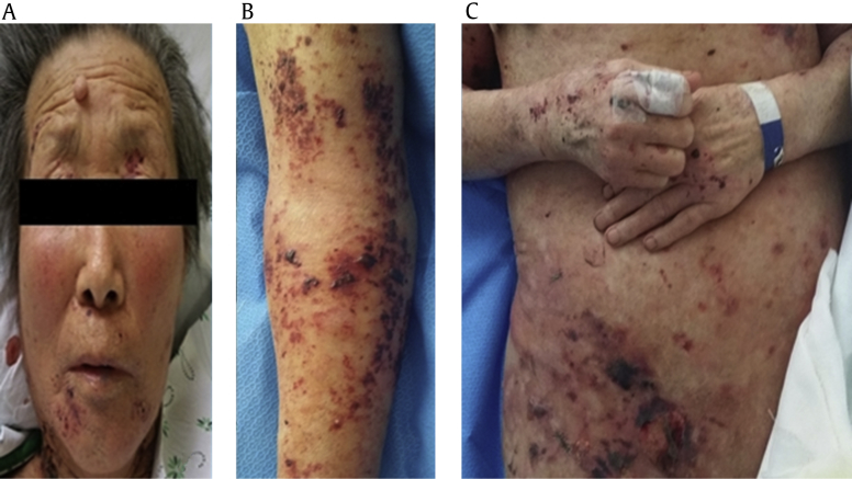
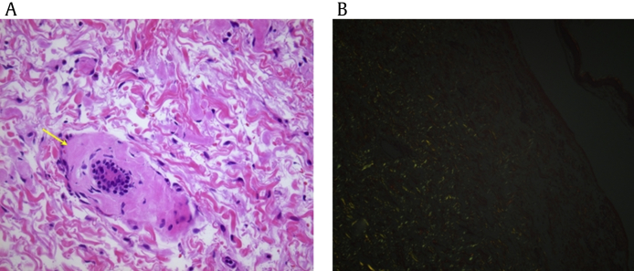
Amyloidosis is a systemic disease involving multiple organs such as heart, kidney, and skin. It can also present with diverse skin lesions such as pinch hemorrhages and maculopapular eruptions; however, hemorrhagic bullous lesions are very rare. Here, I introduce a case of bullous amyloidosis undergoing hemodialysis for the treatment of end-stage renal disease. An 82-year-old woman was diagnosed with chronic kidney disease about 2 years ago. Recently, she visited our clinic for severe uremic symptoms. Laboratory test results showed severe azotemia, nephrotic-range proteinuria, severe anemia, and markedly increased serum-free light chain. Hemodialysis was initiated, and her symptoms were remarkedly relieved. Bone marrow examination and abdominal skin excisional biopsy revealed light-chain lambda-type multiple myeloma and combined amyloid light-chain amyloidosis, respectively. Her multiple myeloma and amyloid light-chain amyloidosis showed a partial response to chemotherapy with cyclophosphamide and prednisolone. However, she developed pneumonia a few weeks after the chemotherapy and was immediately treated with appropriate antibiotics. Several days later, however, she developed extensive hemorrhagic bullous skin lesions without itching sensation on her whole abdomen, back, both extremities, and face (Fig. 1). An excisional biopsy revealed subepidermal blisters with hemorrhage and amyloid deposition. Immunofluorescence staining for IgG, IgA, IgM, C1q, C3, and fibrinogen was negative, whereas Congo red staining for amyloid deposits was positive (Fig. 2).
Figure 1
Hemorrhagic bullous lesions of the patient. Face (A), extremity (B), and trunk (C) show hemorrhagic bullous lesions, which extended to the whole body.

Figure 2
Skin biopsy findings. (A) Hematoxylin–eosin staining shows amorphous eosinophilic nodular deposits and blood vessels (arrow) thickened by eosinophilic materials (original magnification ×400). (B) Congo red staining shows amyloid deposits with apple-green birefringence under polarization microscopy.

- TOOLS
-
METRICS

- Related articles
-
Aspirin Resistance in Patients with End-Stage Renal Disease2011 March;30(2)


 PDF Links
 PubReader
 Full text via DOI
 Download Citation
 Print
Print


