


Introduction
Renal transplantation has become the treatment of choice for suitable candidates with end-stage renal disease [1]. Various urological complications have been reported after herniorrhaphy. Here, we report a case of obstructive uropathy and acute renal failure following inguinal herniorrhaphy with a mesh in a renal transplant patient.
Case report
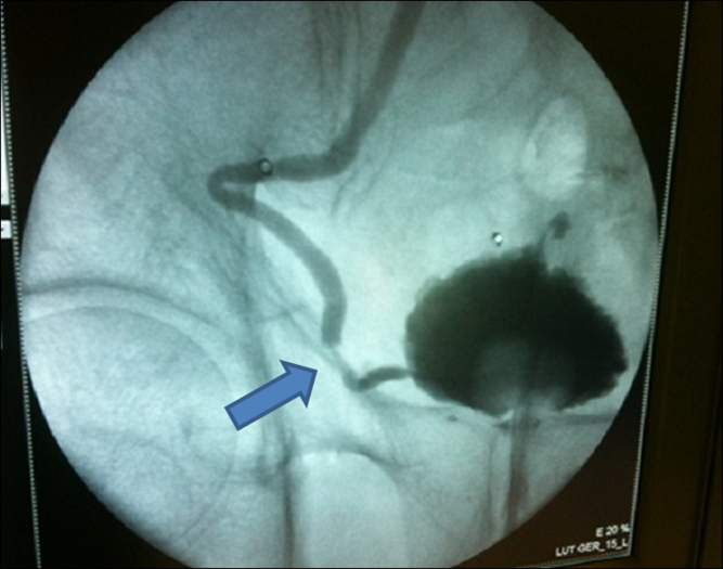
A 67-year-old male patient, who had received kidney transplantation 11 years previously, presented with a right inguinal bulging mass that had been developing for 5 days. He did not have the bulging inguinal mass during the 9-month peritoneal dialysis period before the renal transplantation. He was diagnosed with a right inguinal hernia, in the general surgery department. Before herniorrhaphy, his white blood cell count was 70,900/μL, hemoglobin 10.5 g/dL, platelet count 214,000/uL, serum blood urea nitrogen 24.37 mg/dL, and serum creatinine 1.3 mg/dL. The inguinal hernia was repaired by Prolene Hernia System (PHS) hernioplasty. He presented with severe right lower quadrant pain and oliguria within 24 hours following the operation. Also, his serum creatinine level increased up to 6.46 mg/dL. To identify the cause of the acute renal failure after herniorrhaphy, kidney computed tomography was performed, which revealed a hydronephroureter with distal ureter obstruction. Urgent percutaneous nephrostomy was performed and anterograde pyelography showed the presence of distal ureteral stenosis (Fig. 1). Acute hemodialysis was conducted three times for 4 days, until renal function returned. Subsequently, an operation was performed to remove the PHS mesh. After removal of the mesh, an obstructed ureter was found, which was unfolded. The inguinal hernia was repaired with Lichtenstein herniorrhaphy (hernia repair with the use of mesh). The antegrade ureteral stent (double J stent) insertion was performed. Three days after the repair operation, the patient’s serum creatinine dropped to 1.26 mg/dL from the maximal level 9.05 mg/dL. The double J catheter was removed in 1 month. After the double J stent removal, the patient maintained a stable serum creatinine over the 7-month follow-up.
Discussion
Hernias are common and are diagnosed more frequently in patients undergoing continuous ambulatory peritoneal dialysis [1]. Renal transplant patients often have a previous history of inguinal hernia repairs. Various urological complications have been reported to occur as a result of herniorrhaphy [2]. The bladder and testicular arteries are at risk of injury during routine herniorrhaphy. Among renal transplant patients, reports of urological complications during inguinal herniorrhaphy are rare. During the standard operative procedure for renal transplantation, the kidney graft is placed in the preperitoneal space in the iliac fossa, and the graft ureter is anastomosed to the bladder. Since the ureter of the transplanted kidney is close to the anterior area, there is a risk of ureteral ligation during inguinal herniorrhaphy. Selman et al reported ureteral ligation following inguinal herniorrhaphy in a transplant patient [3]. The Lichtenstein herniorrhaphy has greatly reduced the risk of recurrence of inguinal hernias [4], [5]. The use of mesh plugs has been advocated for decades and has recently achieved acceptance as an appropriate method of hernia repair [1].
We report that ureteral kinking and stenosis (ureteral stenosis) following inguinal herniorrhaphy with the use of a mesh in a transplant patient, could induce hydronephrosis and acute renal failure. This case illustrates hydronephrosis related to the mesh for inguinal hernia repair and demonstrates the need for a safe distance between vital structures and any prosthetic device. Obstructive uropathy should be considered when a renal transplant patient presents with acute renal failure after inguinal herniorrhaphy.
 PDF Links
 PubReader
 Full text via DOI
 Download Citation
 Print
Print


")


